
MS Minute: Emerging Myelin Repair Therapies in Multiple Sclerosis
Remyelination is a promising treatment strategy in multiple sclerosis, with recent clinical trials demonstrating biological efficacy using sensitive visual outcomes and advanced imaging, providing a credible route to promoting neuroprotection and preventing disability.
KEY TAKEAWAYS
- Remyelination therapies may help promote neuroprotection beyond inflammatory disease control.
- VEPs, OCT, MRI, and fluid biomarkers are shaping myelin repair trial design.
- Clemastine, metformin, bexarotene, and other agents are advancing remyelination research.
Multiple sclerosis (MS) is the leading nontraumatic cause of disability in young adults.1 Although highly effective immunomodulatory disease-modifying therapies (DMTs) reduce relapses, progressive disability often continues to develop.2 This ongoing progression, despite successful suppression of central nervous system (CNS) inflammation, demonstrates a therapeutic ceiling for DMTs. New treatment strategies are needed to halt MS progression.
A leading treatment method is to use the nervous system’s endogenous regenerative capacity to remyelinate axons after demyelination.3 Although remyelination occurs naturally, it is insufficient in MS, and its efficiency decreases with advancing age.4 Restoring myelin can reestablish secure nerve conduction, protect underlying axons from degeneration, and potentially improve functional outcomes. This neuroprotective effect positions remyelination as one of the most tractable strategies to prevent disability in MS.
Advances in remyelination biology have identified several therapeutic targets and led to success in phase 2 clinical trials.5-7 These advances have been achieved through the repurposing of existing therapies. As the underlying biology of remyelination failure becomes more clearly defined, novel agents with potential for greater efficacy are entering early-phase clinical development.
In this update, we outline the progression from fundamental remyelination biology to successful clinical trials and highlight the key lessons essential for translating these early successes into routine clinical practice.
Remyelination Biology
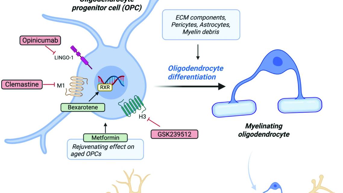
Remyelination is the default regenerative response to demyelination and primarily occurs through the development of newly formed myelinating oligodendrocytes. This process starts with activation of a population of CNS resident stem cells called oligodendrocyte progenitor cells (OPCs), which subsequently divide, migrate to areas of demyelination, and differentiate, resulting in the new oligodendrocytes crucial for effective remyelination.3,8 Multiple cues control this sequence of events, including the inflammatory immune cascade, reactive astrocytes, pericytes, the extracellular matrix, and the demyelinated axons themselves, in addition to intrinsic OPC factors. Due to studies revealing the presence of OPCs surrounding demyelinated lesions, early efforts have focused on targeting the differentiation phase of remyelination.
Two complementary approaches have guided identification of remyelinating compounds. One effective strategy has been phenotypic screening of libraries of US Food and Drug Administration–approved compounds based on functional cellular outcomes. For example, adult OPCs cultured on micropillar assays with high-throughput screening led to identification of M1 antagonists as potential remyelinating drugs, culminating in the repurposing of clemastine in phase 2 trials.9 In parallel, hypothesis-driven approaches have selected drugs based on known cellular targets and pathways implicated in the failure of remyelination in MS (eg, identification of the remyelinating potential of RXR gamma agonism with bexarotene and of AMPK agonism with metformin).10
These advances have been highly productive for effective translation from bench to bedside. However, targeting OPC differentiation alone is unlikely to provide a universal solution across all lesion types or populations. Increasing evidence also shows that oligodendrocytes are morphologically and functionally heterogeneous, influencing remyelination dynamics.11 A more transformative strategy may require harnessing the full regenerative capacity of the oligodendrocyte lineage, including the potential of surviving, mature oligodendrocytes after demyelinating injury.
The Challenge: Measuring Remyelination in Humans
In preclinical studies, remyelination can be histologically identified using high-resolution electron microscopy. However, such approaches cannot be translated to studies in people. Despite this, several neurophysiologic and imaging-based assessments have emerged as sensitive and potentially meaningful outcome measures for clinical trials.

Figure 1: Biology of remyelination and therapeutic targets of recent trials.
Created in BioRender. Moniri, M. (2026) https://BioRender.com/shb8nru
Abbreviations: ECM, extracellular matrix; H3, histamine receptor 3; LINGO-1, leucine-rich repeat and immunoglobulin-like domain-containing protein 1; M1, muscarinic receptor 1; OPC, oligodendrocyte progenitor cell; RXR, retinoid X receptor.
Visual Measures
Because the visual pathway is commonly affected in MS, it provides an accessible, noninvasive, in vivo model for assessing lesion remyelination, and visual pathway biomarkers have therefore become the current standard for detecting myelin repair over the short timescales typical of early-phase clinical trials12 (Figure 1).
Visual Evoked Potentials. Visual evoked potentials (VEPs) appear to be the most sensitive electrophysiologic measure. Generated by the primary visual cortex in response to a visual stimulus, VEPs are most commonly assessed using full-field pattern-reversal paradigms (full-field VEP). Measurement of signal latency and amplitude provides insight into axonal conduction velocity and the numbers of functional fibers. Prolonged latency has been established to be associated with demyelination; it has also been shown that reductions in latency directly follow remyelination (rather than resolution of conduction block or plasticity).13 Consistent with this, changes in VEP latency have demonstrated statistically significant treatment effects across 3 remyelination trials.5-7 However, reliance on VEPs necessitates recruitment of participants with prolonged baseline latency, resulting in high screening failure rates, which exceed 60% in some studies (Table).
Multifocal VEPs offer an alternative approach, stimulating discrete regions of the visual field, enabling regional changes to be assessed. Multifocal VEPs appeared to be more sensitive in a substudy of opicinumab In Acute Optic Neuritis (RENEW; NCT01721161),14 but this result was not replicated in our recent trial of metformin and clemastine (Cambridge Centre for Myelin Repair Trial Two [CCMR Two]; NCT05131828), in which full-field VEP outperformed multifocal VEP.6

Optical Coherence Tomography. Optical coherence tomography (OCT) is a rapid and noninvasive measure that can complement VEP through quantification of axonal integrity in retinal layers, most commonly in the peripapillary retinal nerve fiber layer (RNFL) and the macular ganglion cell–inner plexiform layer.15 Cell bodies of retinal ganglion cells lie in the ganglion cell layer and their axons are found in the RNFL, reflecting neuronal and axonal integrity in MS. OCT does not directly detect myelin loss,16 with myelination starting behind the lamina cribrosa. OCT is therefore best interpreted as a measure of neuroaxonal loss secondary to demyelination. This makes OCT suited to capturing long-term neuroprotective effects, but it is more commonly used as a screening tool to ensure sufficient axonal scaffolding for remyelination; therefore, many trials exclude participants with a global peripapillary RNFL thickness <70 μm.6,7
Eye Movement Abnormalities. Eye movement abnormalities represent a further functional approach to test remyelination in MS. High-frequency infrared eye tracking enables precise quantification of reaction times and of interocular dysconjugacy,17 allowing the detection of internuclear ophthalmoplegia (INO) caused by lesions of the medial longitudinal fasciculus. Because this heavily myelinated tract is often demyelinated in people with MS,18 improvement of INO can be used to measure remyelination.17 However, this requires the presence of an INO measure at baseline, again resulting in a high screening failure rate comparable to trials using VEP latency.
Low-Contrast Letter Acuity. Low-contrast letter acuity (LCLA) is arguably the most clinically meaningful visual outcome and has been deployed across several clinical trials6,19 (Table). However, LCLA lacks specificity to myelin repair, is susceptible to learning effects, and is unlikely to change over short trial durations.
Imaging and Biomarkers
Visual outcome measures provide sensitive, low-cost, noninvasive tools for assessing remyelination. Nevertheless, heavy reliance on a single functional system introduces pathway-specific bias. For robust evaluation of remyelination, visual biomarkers should be integrated with complementary measures from magnetic resonance imaging (MRI) and potentially fluid biomarkers (Figure 2).
Myelin Imaging. MRI is central to the diagnosis and management of MS, yet conventional clinical MRI sequences are nonspecific to myelin content. Advanced sequences, including myelin water fraction, diffusion tensor imaging, and magnetization transfer ratio (MTR), have been used to gain insight into remyelination through quantification of myelin water, water diffusion, and macromolecular content. Clemastine treatment was associated with improvements in corpus callosum myelin water fraction.20 After bexarotene treatment, remyelination was demonstrated by increases in whole-lesion MTR in gray matter lesions5 and through voxel-level analyses of white matter lesions.21 However, these techniques provide indirect assessments of myelination and are susceptible to confounding pathologic processes. Positron emission tomography offers a highly myelin-specific alternative,22 but limited availability and radiation exposure remain barriers to its use as a clinical trial outcome measure.
Blood Biomarkers. One of the most accessible blood biomarkers is neurofilament light chain (NfL), a structural component of the neuronal cytoskeleton. Elevated serum NfL has been widely accepted as a sensitive marker of inflammatory disease activity.23 Encouraging data indicate that remyelination induced by both bexarotene and clemastine is associated with reductions in serum NfL, providing early evidence that successful remyelination may stabilize axonal integrity. However, NfL remains an indirect readout of remyelination and, despite its scalability, is likely to lack sensitivity to modest or spatially restricted myelin repair. Looking ahead, a more informative strategy may be the identification of additional CNS-derived proteins released during active remyelination that can be detected in blood, enabling scalable, mechanism-specific biomarkers to directly capture myelin repair in clinical trials.

Figure 2. Common outcome measures in remyelination trials.
Schematic overview of structural and functional outcome measures used to assess remyelination along the visual and oculomotor pathways.
Created in BioRender. Moniri, M. (2026) https://BioRender.com/8ixe5s9
Abbreviation: VEP, visual evoked potential.
Recent Remyelination Trials
Recent remyelination trials mark the progression toward a new class in MS therapeutics. Most contemporary strategies aim to enhance OPC differentiation. Therapeutic targets have included antagonism of muscarinic M1 receptors (eg, clemastine, PIPE-307), inverse agonism of H3 receptors (eg, GSK239512), agonism of retinoic acid X receptors (eg, bexarotene), and selective estrogen receptor modulation (eg, bazedoxifene), with variable effects on remyelination (Table).
For instance, following the phase 2 Cambridge Centre for Myelin Repair One (CCMR One) trial of bexarotene, which failed to meet its primary end point but showed clear effects on remyelination through reductions in VEP latency and improvements in MTR of gray matter lesions and sublesional components of white matter lesions, we changed our approach in the CCMR Two trial of metformin and clemastine. In CCMR Two, change in VEP latency was our primary outcome measure, and participants with chronic stable optic neuropathy were recruited. This design showed a positive effect, despite a smaller effect size than that seen in the original CCMR One. This trial validates the use of a model system (such as the visual pathway) to obtain a go/no-go signal of biological efficacy. Demonstrating a clinically significant improvement in a measure such as the Expanded Disability Status Scale or visual acuity, particularly in individuals with stable MS, would be challenging over a short-duration clinical trial; recent trials have failed to deliver positive results using LCLA as a primary readout.28
Phase 2 trials should first establish biological evidence of efficacy before progressing to longer-term trials (with outcomes of disability or visual function). The timescale for a clinically significant effect from remyelination has not been determined but is likely to extend several years. In the short term, it is essential to follow up participants from previous remyelination trials to capture any long-term benefits from treatment with remyelinating drugs, even if used for short durations (as we showed with bexarotene29).
Emerging Approaches
Beyond these current strategies, emerging approaches to target remyelination through alternative and potentially complementary mechanisms are being investigated. Neural stem cell transplantation has been shown to enhance remyelination of lesions in preclinical models,30 and phase 1 trial data have demonstrated safety in people with progressive MS.31 Nanoparticle-based approaches have also been investigated, with nanocrystalline gold promoting remyelination in preclinical models,32 although published results from completed clinical trials are pending. In parallel, there is interest in physiologic modulators of repair, with evidence suggesting that increased activity levels, such as exercise, may enhance OPC proliferation.33 These developments highlight the rapid evolution in this area of research.
Conclusions
Remyelination represents one of the most accessible strategies for achieving neuroprotection in MS. Demonstrating remyelination, however, remains a challenge. Early-phase trials require highly sensitive outcome measures, such as changes in VEP latency, but the timescale over which remyelination translates into clinically meaningful improvements may extend well beyond the duration of typical studies. The widespread use of standard-of-care DMTs is also likely to attenuate observable differences between treatment and control groups, further complicating efforts to provide evidence of the additional benefit of remyelination on downstream neuroprotection.
In response, emerging approaches, such as eye tracking, lesion-specific and quantitative MRI analyses, and increasingly sensitive blood-based biomarkers, are being integrated with established visual electrophysiology to deliver multimodal, functionally meaningful readouts of myelin repair. Longer-term studies with extended follow-up will be essential to define the temporal relationship between remyelination and durable functional benefit.
Careful consideration of patient selection, disease stage, age, and outcome measure choice remains central to optimizing trial design. Advances in both therapeutic efficacy and measurement sensitivity are required, but phase 2 trials must first establish biological remyelination, grounded in the expectation that successful myelin repair will ultimately confer long-term neuroprotection, even if the clinically meaningful benefits only become apparent over several years.
Ready to Claim Your Credits?
You have attempts to pass this post-test. Take your time and review carefully before submitting.
Good luck!
Recommended
 MS & Immune Disorders
Autologous Stem Cell Transplantation Inducing Durable Remission in Relapsing Tumefactive Multiple Sclerosis
Andrea Waser, BBiomedSc, MBBS; Mike Boggild, MD, FRACPAndrea Waser, BBiomedSc, MBBS; Mike Boggild, MD, FRACP- MS & Immune Disorders
MS Minute: Chimeric Antigen Receptor T-Cell Therapy for Progressive Multiple Sclerosis: Rationale, Evidence, and Therapeutic Potential
Sarah Conway, MD; Kristin Galetta, MD, MSeDSarah Conway, MD; Kristin Galetta, MD, MSeD - Imaging & Testing
Practical MRI in Neurology: Use Cases, Technologies, and Opportunities for Outpatient Clinics
Joseph Fritz, PhD; Edmond A. Knopp, MD, DABR, FASFNRJoseph Fritz, PhD; Edmond A. Knopp, MD, DABR, FASFNR