
Autologous Stem Cell Transplantation Inducing Durable Remission in Relapsing Tumefactive Multiple Sclerosis
Autologous stem cell transplantation may be an effective escalation strategy for highly active tumefactive multiple sclerosis refractory to high-efficacy disease-modifying therapies.
KEY TAKEAWAYS
- Tumefactive multiple sclerosis can create diagnostic and therapeutic challenges when large inflammatory lesions resemble other neurologic disease processes.
- Highly active disease may continue despite multiple conventional and high-efficacy treatment strategies.
- This case highlights the clinical considerations surrounding escalation therapy in a rare and difficult-to-treat form of multiple sclerosis.
Case Presentation
AB, aged early 20 years, presented to the emergency department with a 3-day history of left-sided visual disturbance and progressive right-sided weakness. There were no associated headaches, fevers, weight loss, or other constitutional symptoms. The medical history was unremarkable, with no regular medications. AB reported a smoking history of 5 cigarettes daily, minimal alcohol intake, and no illicit substance use.
Neurologic examination revealed marked right-sided weakness affecting both upper and lower limbs. Right upper limb grip strength was preserved (5/5), with reduced elbow flexion (4/5) and extension (3/5). Right lower limb hip flexion was reduced (3/5), with marked weakness affecting knee extension (2/5), knee flexion (1/5), ankle plantar flexion (2/5), and dorsiflexion (0/5). Tone and deep tendon reflexes were initially normal, with downgoing plantars and no clonus or Hoffmann sign. Sensory examination revealed reduced light and sharp touch sensation in the right lower limb.
Over the course of admission, AB developed increased tone, hyperreflexia, and clonus in the right limbs, consistent with evolving upper motor neuron signs.1 Visual field assessment demonstrated a left homonymous hemianopia. The remainder of the neurologic examination was unremarkable.
AB was admitted under the neurosurgical department, with initial management including intravenous dexamethasone 8 mg, then 4 mg twice daily. Seizure prophylaxis was also initiated with 1 g levetiracetam, followed by 500 mg twice daily.
Diagnostic Process
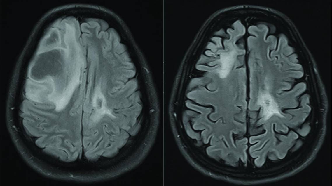
Initial investigations, comprising a full blood count, metabolic panel, coagulation studies, and hematinics, were unremarkable. CT brain scan with contrast revealed an incomplete rim-enhancing lesion within the left frontal-parietal lobe with surrounding vasogenic edema and mild mass effect. CT of the chest, abdomen, and pelvis with contrast revealed no evidence of a primary mass lesion or metastatic deposits. A whole-body PET scan revealed photopenia in the left frontal cortex secondary to perilesional edema, with no evidence of an extracranial primary site of malignancy. MRI of the brain and spine demonstrated large, peripherally enhancing intra-axial lesions located in the left frontal and right occipital lobes (Figure 1A). A T2-hyperintense cord lesion was noted at the level of the C6 vertebra, with no postcontrast enhancement.

Figure 1. Axial T2-weighted fluid-attenuated inversion recovery MRI brain scans before and after autologous stem cell transplantation. In January 2022, at the first clinical and radiologic relapse, a large new lesion within the right central frontal lobe displays diffuse high T2-weighted and fluid-attenuated inversion recovery signal, with a reduction in the previously identified left frontal lobe demyelinating lesion compared with the initial presentation in December 2021 (A). In December 2024, 21 months after autologous stem cell transplantation, a reduction in established demyelinating lesions is evident, with no new demyelinating lesions seen (B).
Following this initial workup, a stereotactic biopsy of the left frontal lesion was obtained by the neurosurgical team on day 4 of admission. Silver and myelin staining revealed widespread myelin degeneration with relative preservation of axons. Flow cytometric analysis on the tissue sample showed no abnormal B- or T-cell populations to suggest lymphoma; however, AB had received 4 days of dexamethasone at the time of biopsy. These histopathologic findings were consistent with tumefactive demyelination, and a diagnosis of tumefactive multiple sclerosis (TMS) was established.
Following diagnosis of TMS from the tissue biopsy, the neurology department was consulted and assumed ongoing care of AB. Further investigations for demyelinating conditions were completed. Neuronal antibody testing (anti–myelin oligodendrocyte glycoprotein and anti–neuromyelitis optica immunoglobulin G antibodies) was negative for neuromyelitis optica spectrum disorder and myelin oligodendrocyte glycoprotein antibody disease. Cerebrospinal fluid analysis was unremarkable and specifically notable for the absence of oligoclonal bands. This finding, although atypical in multiple sclerosis (MS), is not unexpected in TMS, in which oligoclonal bands are reported in ~50% of individuals presenting with tumefactive demyelination.2
In light of the definitive radiologic and histopathologic findings, further ancillary investigations, such as visual evoked potentials and optical coherence tomography, were not completed, as they were not expected to alter further management.
Case Resolution
Over the course of 15 months, AB was admitted on 6 further occasions. Three of these presentations represented clinical and radiologic relapse, characterized by new symptoms and newly identified enhancing lesions on MRI. The remaining 3 presentations were marked by progression of existing symptoms without imaging evidence of new demyelinating activity.
Multiple lines of conventional and disease-modifying therapy failed to induce durable remission. These included several courses of high-dose intravenous steroids, 3 courses of therapeutic plasma exchange, and sequential trials of 3 high-efficacy disease-modifying therapies (HETs): natalizumab (January 2022), alemtuzumab (April 2022), and ocrelizumab (Ocrevus; Genentech, South San Francisco, CA; November 2022). AB’s Expanded Disability Status Scale (EDSS) score worsened with each relapse, reaching 6 at the sixth presentation (Figure 2), by which point an axillary crutch was required for mobilization due to increasing gait limitation.

Figure 2. Expanded Disability Status Scale (EDSS) score evolution.
As salvage therapy, a high-dose cyclophosphamide and thymoglobulin autologous stem cell transplantation (ASCT) was completed in March 2023. Before ASCT, AB had a 3-month washout period from HET, during which time steroids were used as a temporizing measure during a further clinical relapse. AB underwent stem cell mobilization with cyclophosphamide 2 g/m2 with daily filgrastim injections at 10 μg/kg starting on day 3 of mobilization. Stem cell harvest through leukapheresis was successfully completed on day 9 of mobilization. Cryopreservation of stem cells was completed as per Australian laboratory standards in 10% dimethyl sulfoxide and rapidly thawed at the bedside for reinfusion. The transplant protocol included cyclophosphamide 50 mg/kg on day −5 through day −2 and antithymocyte globulin (rabbit) 0.5 mg/kg on day −5 through day −1. The treatment was tolerated without any clinically significant complications other than expected transient hematologic toxicity. This included an episode of culture-negative febrile neutropenia without sepsis, for which AB received 5 days of intravenous piperacillin-tazobactam. In addition to this, AB experienced cytopenias requiring transfusion with 5 units of packed red cells and 5 units of platelets in total between days −2 and 11 of ASCT.
After ASCT, the EDSS score improved from 6 to 3 (see Figure 2 in the article on www.practicalneurology.com). AB has remained in remission for >2 years posttransplant, with no further clinical or radiologic relapse. Persisting neurologic deficits include an ongoing left homonymous hemianopia and a mild right foot drop that does not affect mobility. Follow-up MRI performed at 3, 8, and 21 months posttransplant has demonstrated no new active lesions to date (Figure 1B).
Discussion
TMS, a rare variant of MS, is characterized by large, inflammatory, tumor-like lesions within the central nervous system (CNS).3 It is thought to represent <1% of all MS cases and poses considerable therapeutic challenges, as conventional MS therapies may lack comparable efficacy in TMS.4 Historically, therapeutic approaches have resembled those used in conventional MS, including high-dose corticosteroids, therapeutic plasma exchange, and HET. However, evidence supporting the efficacy of these strategies in TMS remains limited, with no comparative outcome data to inform management.5
An important limitation when extrapolating the efficacy of HET in TMS is the underrepresentation of participants with tumefactive lesions in pivotal MS trials. Major studies supporting HET—including Safety and Efficacy of Natalizumab in the Treatment of Multiple Sclerosis (AFFIRM; NCT00027300), Comparison of Alemtuzumab and Rebif Efficacy in Multiple Sclerosis (CARE-MS; NCT00530348 [CARE-MS I] and NCT00548405 [CARE-MS II]), A Study of Ocrelizumab in Comparison With Interferon Beta-1a in Patients With Relapsing Multiple Sclerosis (OPERA; NCT01247324 [OPERA I] and NCT01412333 [OPERA II]), and Ofatumumab Versus Teriflunomide in Patients With Relapsing Multiple Sclerosis (ASCLEPIOS; NCT02792218 [ASCLEPIOS I] and NCT02792231 [ASCLEPIOS II])—enrolled participants with an established diagnosis of relapsing MS and MRI findings consistent with MS. Individuals presenting with tumefactive demyelinating lesions may not fulfill these diagnostic criteria at presentation, given that radiologic appearances may mimic primary CNS malignancy, lymphoma, or infection, and often necessitate additional investigations, including biopsy, before a definitive diagnosis of demyelination can be established. This diagnostic ambiguity represents an important source of selection bias and limits the generalizability of HET trial data to people with TMS. Therefore, their efficacy in TMS remains uncertain and is supported primarily by observational data and case reports.5
A further consideration in TMS is the reported paradoxical or rebound emergence of tumefactive demyelinating lesions, seen to occur both during treatment as well as discontinuation of natalizumab and sphingosine-1-phosphate receptor modulators. This phenomenon is thought to reflect altered lymphocyte trafficking with subsequent reentry of lymphocytes into the CNS.6 AB commenced natalizumab therapy in January 2022 and received 3 doses before discontinuation due to a rising JC virus antibody titer. In April 2022, AB developed a new left occipital lesion. Given the close temporal relationship to natalizumab withdrawal, it is possible that a paradoxical response may have contributed to this episode. However, the persistence of disease activity despite sequential treatment with alternative HETs (alemtuzumab and ocrelizumab) suggests that the overall disease course was driven by treatment-refractory TMS rather than a discrete natalizumab-related rebound event.
ASCT is gaining increasing traction in the therapeutic landscape of MS. To date, only 2 randomized controlled trials (RCTs) have been completed, with 3 additional studies currently underway in the United Kingdom (Autologous Hematopoietic Stem Cell Transplantation Versus Best Available Disease-Modifying Therapy for Highly Active Relapsing Multiple Sclerosis [STAR-MS]; ISRCTN88667898), United States (Best Available Therapy Versus Autologous Hematopoietic Stem Cell Transplant for Treatment-Resistant Relapsing Multiple Sclerosis [BEAT-MS]; NCT04047628), and Norway (Randomized Autologous Hematopoietic Stem Cell Transplantation Versus Alemtuzumab, Cladribine, or Ocrelizumab for Relapsing-Remitting Multiple Sclerosis [RAM-MS]; NCT03477500).7-9 A 2019 RCT involving 110 participants with relapsing-remitting MS compared ASCT with continued standard therapy. The study demonstrated a significantly lower rate of disease progression at 5 years in the ASCT group (9.7%) compared with the control group (75.3%). One-year EDSS scores were also improved following ASCT. Median time to progression was 24 months in the DMT group but was not reached in the ASCT group because of the low number of progression events.8 In addition to this impressive albeit limited data regarding the efficacy of ASCT, retrospective research has consistently demonstrated relative safety, with treatment-related mortality for people undergoing ASCT reported at ~0.3%.9 Existing RCTs, however, provide limited evidence on the efficacy of ASCT relative to contemporary HET, given that their comparator arms do not adequately represent the modern HET landscape.8
A 2025 expert consensus statement developed by the European Committee for Treatment and Research in Multiple Sclerosis and the European Society for Blood and Marrow Transplantation outlined a series of evidence-based recommendations regarding MS treatment. The consensus supports the broader adoption of ASCT as an escalation therapy for individuals with highly active MS who have failed HET, emphasizing the importance of early referral for evaluation.10 This case describes failure of multiple HETs in an individual with highly active, treatment-refractory TMS, followed by sustained clinical and radiologic remission after ASCT. Whereas a single case cannot establish treatment efficacy, this outcome, in conjunction with the European Committee for Treatment and Research in Multiple Sclerosis/European Society for Blood and Marrow Transplantation consensus recommendations, supports consideration of ASCT as an escalation strategy in highly active TMS. This case presentation also highlights the need to include tumefactive variants in future research to better inform treatment decisions for this rare presentation.
Ready to Claim Your Credits?
You have attempts to pass this post-test. Take your time and review carefully before submitting.
Good luck!
Recommended
 MS & Immune Disorders
MS Minute: Emerging Myelin Repair Therapies in Multiple Sclerosis
Mahtab Moniri, MPhil; Meher Sabharwal, BA (Hons)Mahtab Moniri, MPhil; Meher Sabharwal, BA (Hons)- Alzheimer Disease & Dementias
Special Report: 2025 Neurology FDA Drug & Device Approvals & Clearances