
The New Era of Deep Brain Stimulation Programming in Parkinson Disease
Directional stimulation, image-guided programming, neural sensing, adaptive DBS, and remote programming are making deep brain stimulation programming more precise and personalized.
KEY TAKEAWAYS
- New DBS technologies are giving clinicians more ways to personalize stimulation while reducing reliance on traditional trial-and-error programming.
- Neural sensing and imaging-based tools are expanding the information available to guide Parkinson disease treatment decisions.
- As DBS becomes more responsive and data-driven, clinicians must balance technologic capabilities with patient-specific goals and clinical expertise.
Deep brain stimulation (DBS) is a Food and Drug Administration–approved treatment for medication-refractory Parkinson disease (PD), essential tremor, dystonia (under a Humanitarian Device Exemption), obsessive-compulsive disorder, and focal epilepsy. It was introduced clinically in 1987 for the treatment of essential tremor, building on earlier research into electrical stimulation of the brain.1 Subsequently, numerous randomized controlled trials have demonstrated the safety and efficacy of DBS, establishing it as a powerful and widely accepted therapeutic option for multiple movement disorders.
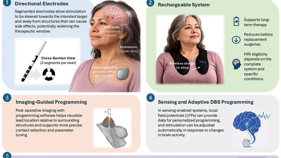
Advances in hardware and software are increasingly transforming clinical DBS practice. Newer DBS systems, featuring rechargeable batteries, MRI compatibility, and advanced lead designs, provide greater flexibility in spatially directing current and selecting a broader range of stimulation settings. Image-guided programming tools support more precise contact selection and parameter tuning, reducing trial-and-error methods. With sensing-enabled DBS systems, clinicians can identify regions within target nuclei that most closely correspond to specific symptoms, monitor local field potentials (LFPs) in real time, and adjust stimulation based on patient-specific neural signatures. Remote and teleprogramming capabilities are expanding access for patients who live far from DBS centers, enabling more continuous care. Together, these technologies streamline workflow, improve personalization of therapy, and enhance clinical outcomes in routine DBS practice (Figure).
Traditional Approach
DBS programming typically begins once lead placement is confirmed and hardware integrity is verified with impedance checks. To avoid confounding effects from microlesion phenomena, such as acute motor improvements caused by local tissue manipulation, microhemorrhages, or edema, programming is usually initiated 3 to 4 weeks after implantation.
DBS programming focuses on optimizing 4 key stimulation parameters. Contact configuration determines which electrode contacts are activated and the direction and polarity of current delivery. Stimulation amplitude or current determines the strength of electrical stimulation delivered to the target. Pulse width determines the duration of each electrical pulse and influences the neural elements recruited by stimulation. Frequency determines the number of pulses delivered per second and can influence both therapeutic benefit and stimulation-related side effects.2
Traditional programming, which relies on trial and error to identify optimal contacts and stimulation parameters, is time-consuming and requires considerable expertise. This approach may lack precision, occasionally deliver excessive stimulation, and cause adverse effects, such as dysarthria, freezing, or stimulation-induced dyskinesia. In-clinic programming sessions may capture acute adverse effects but may not identify those that emerge with chronic stimulation.
Hardware Advances
Recent advancements aim to deliver a precise, personalized, and optimized stimulation dose for efficacy while minimizing side effects.3 Electrical fields can now be steered horizontally or vertically along the lead. Eight-contact directional DBS leads feature segmented contacts at the middle levels arranged in a circular formation, allowing programmable axial steering of current for more precise control and improved therapeutic outcomes. Directional programming is typically used when the threshold for stimulation-related side effects is low or when the therapeutic window—the range of currents producing clinical benefits with minimal adverse effects—is <2 mA.
In such cases, programmers often use segment mode, which delivers current directionally through individual segments, rather than ring mode, in which current is emitted omnidirectionally through all 3 segments. Transitioning from ring to segment mode requires caution, as smaller electrodes result in higher charge density, necessitating finer intensity adjustments (0.1–0.3 mA per step versus the traditional 0.5 mA) and limiting current to approximately 3.4 mA per contact.3
Activating multiple adjacent segmented electrodes may reduce directionality because current can spread through electrode edges, creating a broader field than the electrode surface itself. Directional systems have been shown to lower the current required for clinical benefit while increasing the side-effect threshold, with studies demonstrating an approximately 40% reduction in therapeutic current and a similar increase in the therapeutic window.4 Expanded pulse width ranges and adjustable frequencies allow more tailored treatment for both appendicular and axial symptoms. Clinical studies demonstrate that short pulse widths, directional steering, or interleaved stimulation can improve speech intelligibility; reduce dysarthria, dyskinesia, and pyramidal side effects; and maintain effective motor symptom control over months.5
Rechargeable DBS systems use implanted batteries that can be externally recharged, reducing the need for frequent replacement surgeries, improving patient convenience, and lowering health care costs.
Software Advances
Image-Guided Programming
Image-guided programming integrates neuroimaging with DBS by visualizing electrode location relative to target nuclei and surrounding networks to further aid in spatial precision. Fusion of postoperative CT or MRI with preoperative imaging enables identification of active contacts and estimation of the volume of tissue activated, supporting more precise and efficient programming. Visualization platforms such as SureTune (Medtronic; Minneapolis, MN), STIMVIEW-XT (Boston Scientific; Marlborough, MA), and Illumina 3D (Illumina; San Diego, CA) generate patient-specific, 3-dimensional models of lead trajectories and anatomy, reducing trial and error, shortening programming time, and minimizing stimulation-induced side effects.3

Figure. Contemporary deep brain stimulation (DBS) technology. Advances include directional electrodes for more precise current steering, rechargeable systems, image-guided programming, sensing-enabled and adaptive DBS programming, and remote programming capabilities. Abbreviations: IPG, implantable pulse generator; LFP, local field potential. Illustration generated with ChatGPT 5.1 (OpenAI, San Francisco, CA) and edited and illustrated by Kimmy Su, MD, PhD.
These technical advances are now increasingly available to clinicians and offer clear advantages, particularly in improving the spatial precision of DBS programming. However, their integration into routine clinical practice may present challenges. For example, some academic and health care institutions may have cybersecurity and data privacy concerns related to connected DBS technologies. From a technical perspective, tissue inhomogeneity may alter current distribution and complicate the interpretation of neural signals, and impedance-related factors may influence stimulation delivery and sensing measurements. In addition, lead migration over time may affect the accuracy of imaging-based targeting and the relationship between the programmed stimulation field and the intended neural structures. Connectome-guided targeting (mostly used in research settings) integrates diffusion tractography and functional imaging and enables delineation of structural and functional connectivity to identify neural networks associated with therapeutic benefit and stimulation-related adverse effects.
LFP-Guided Programming
LFPs are thought to primarily reflect the summed excitatory and inhibitory postsynaptic currents within a volume of neural tissue surrounding the DBS electrode, with relatively little contribution from action potentials of individual neurons. Their characteristics are influenced by multiple factors, including neuronal and synaptic sources, the size of the recording volume, electrode geometry and surface properties, and electrode–tissue interface impedance.
Advances in sensing-enabled DBS technology now allow reliable in-clinic and at-home monitoring of LFPs, opening the door to more precise, physiology-informed stimulation delivery. Clinically available systems such as Percept PC and RC (Medtronic) use implanted stimulation electrodes to record LFPs in real time, providing insights into disease-related neural dynamics. A substantial body of evidence links specific frequency bands to clinical motor states. Beta-band activity (13–30 Hz) correlates strongly with bradykinesia and rigidity, and reductions in beta power after levodopa or DBS are commonly used to guide dose titration and programming. Theta (4–8 Hz) and alpha (8–12 Hz) band activity are associated with tremor, although these low-frequency oscillations may also appear in other conditions, such as tics or levodopa-induced dyskinesias. Higher-frequency gamma activity (30–90 Hz) has been linked to dyskinesias, and emerging data indicate that finely tuned gamma activity correlates with levodopa-induced dyskinesia, particularly severity.6
The ability to objectively sense LFPs over the long term provides several advantages, including improved characterization of disease states and traits, monitoring medication “on”–“off” cycling and circadian influences, identifying potential issues with medication adherence, and guiding optimal contact selection during programming. However, sensing technology also has important limitations. LFP peaks are not always disease- or symptom-specific; signal interpretation can be confounded by stimulation artifacts, movement, or electrocardiographic contamination; and device sampling rates may limit spectral resolution. Continuous sensing may increase battery consumption, although rechargeable systems can partially mitigate this issue.
In 2025, adaptive deep brain stimulation (aDBS) received regulatory approval based on evidence from the Adaptive DBS Algorithm for Personalized Therapy in Parkinson’s Disease trial (ADAPT-PD; NCT04547712). Unlike conventional DBS, aDBS dynamically adjusts stimulation in real time based on neural biomarkers, most commonly beta-band activity, adding temporal precision to the spatial precision provided by directional stimulation. ADAPT-PD demonstrated that aDBS could provide motor control comparable with conventional DBS while reducing stimulation delivery and energy consumption, with potential reductions in stimulation-related adverse effects.7 Current evidence is primarily from PD, and aDBS may be particularly useful for individuals with motor fluctuations, tremor, or dyskinesias. Longer-term studies are needed to determine its broader clinical applicability and optimal programming strategies.
Remote Programming
Remote DBS programming uses telemedicine platforms to allow clinicians to adjust settings without requiring in-person visits, improving access for people living in remote areas, with mobility limitations, or with limited caregiver support. Clinicians can assess symptoms, modify stimulation parameters, and check device impedance and battery status remotely. Individuals may also be able to make minor adjustments or switch between preset programs within clinician-defined limits.
Advanced systems, such as NeuroSphere Virtual Clinic (Abbott; Abbott Park, IL), enable full remote programming with strong security and patient-controlled access. While remote programming reduces travel burden and cost, improper self-adjustment may delay optimization and increase side effects, highlighting the importance of patient education, reliable internet connectivity, privacy, and structured preparation for sessions to ensure safety and optimal outcomes.3
Automated Programming
Automated programming uses computational algorithms that integrate imaging, clinical data, and machine learning to guide stimulation parameter selection. By analyzing lead location, tissue activation, and symptom response, these approaches can reduce programming time and improve consistency while maintaining clinical effectiveness. For example, the StimFit algorithm (MATLAB; Natick, MA) uses imaging-derived and clinical data to predict optimal settings and demonstrated motor outcomes noninferior to conventional programming used for PD. However, limitations such as incomplete modeling of tissue variability and dynamic clinical states highlight the continued need for clinician oversight and further validation.
Conclusion
DBS programming is evolving from an empirical, trial-and-error process toward a more precise, personalized, and data-driven approach. Advances in hardware, including directional leads and rechargeable systems, have improved the spatial precision and flexibility of stimulation, while image-guided programming, neural sensing, and connectomic approaches provide increasingly objective information to guide treatment. Adaptive DBS further enables stimulation to be dynamically adjusted according to neural activity, while remote and automated programming may improve access, efficiency, and consistency.
Together, these advances are moving DBS toward precision neuromodulation, integrating patient-specific anatomy, connectivity, neural physiology, and clinical states. Despite remaining challenges—including biomarker specificity, tissue variability, sensing artifacts, cybersecurity, and the need for broader validation—technology is poised to increasingly complement clinical expertise and transform DBS into a more individualized and responsive therapy.
Ready to Claim Your Credits?
You have attempts to pass this post-test. Take your time and review carefully before submitting.
Good luck!
Recommended
 Movement Disorders
Adaptive Deep Brain Stimulation for Parkinson Disease: Patient Selection, Programming, and Practical Considerations
Pravin Khemani, MDPravin Khemani, MD- SEP-OCT 2026 ISSUE
Wearable Devices in Parkinson Disease: A Practical Guide for Clinicians
Siegfried Hirczy, MD; Ruth B. Schneider, MDSiegfried Hirczy, MD; Ruth B. Schneider, MD - SEP-OCT 2026 ISSUE
Assistive Technology for Communication in Movement Disorders
Nicholas A. Schuck, MDNicholas A. Schuck, MD