
Neural Antibodies in Seizures With a Suspected Autoimmune Etiology: Testing, Interpretation, and Clinical Significance
A seizure-centered approach can help clinicians identify patients with a possible autoimmune etiology, select appropriate antibody testing, interpret the results, and understand their long-term significance, including during relapses.
KEY TAKEAWAYS
- Antibody-associated seizures may present in the context of acute or subacute encephalitis, new-onset refractory status epilepticus (NORSE), febrile infection-related epilepsy syndrome (FIRES), or an insidious, often drug-resistant seizure disorder without overt encephalitic features.
- Neural antibody testing should be guided by clinical phenotype, with autoimmune encephalitis diagnostic criteria and the Antibody Prevalence in Epilepsy and Encephalopathy 2 score (APE2) helping to identify individuals for testing.
- Paired serum and cerebrospinal fluid panel testing is preferred because antibody sensitivity and specificity differ by specimen and assay, and different antibodies may have overlapping clinical phenotypes.
- A positive antibody result alone does not establish autoimmune central nervous system disease; a compatible clinical phenotype is required, and interpretation should consider the specimen tested, assay method, antibody concentration, and need for confirmatory testing.
About half of focal seizure disorders have an unknown etiology1 and 3.4% may have an autoimmune cause associated with antibodies.2 Identifying individuals with an autoimmune etiology is important because seizures may be resistant to antiseizure medications (ASMs) alone,3,4 and early immunotherapy is associated with better outcomes.4-7 Conversely, indiscriminate testing risks finding clinically irrelevant antibodies, a common cause of misdiagnosis in autoimmune encephalitis (AE).8 Thus, the clinical value of neural antibody testing depends not only on identifying whom to test, but also on selecting the appropriate specimen and assay and interpreting results in the context of the clinical phenotype.
This article reviews a seizure-centered approach to determining who warrants antibody testing, selecting appropriate specimens and assays, interpreting positive and negative results, and determining the significance of antibodies after diagnosis.
Identifying Individuals for Antibody Testing: Encephalitic vs Isolated Seizure Presentations
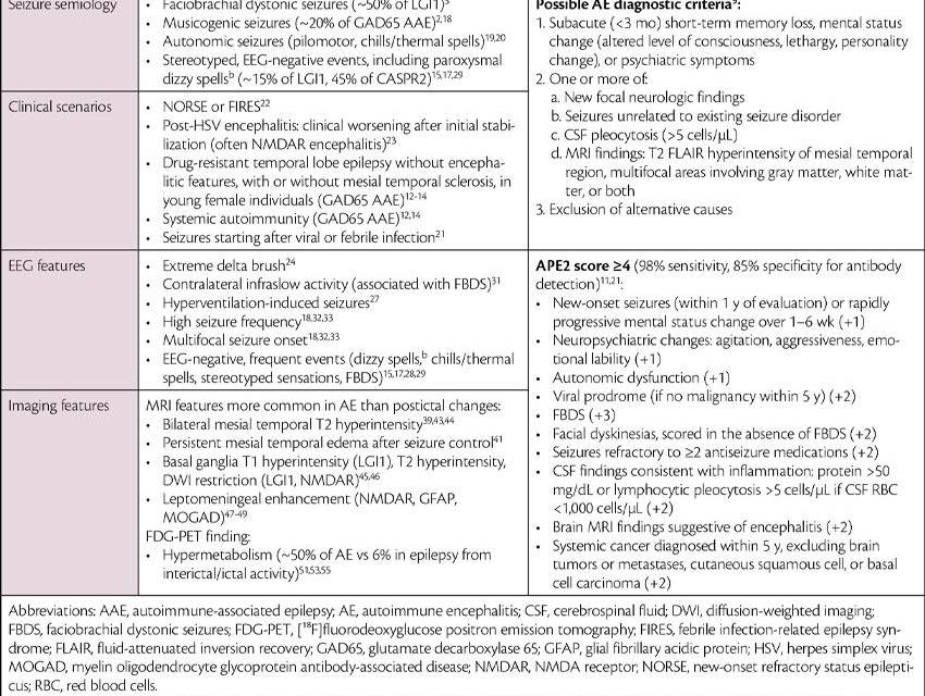
Individuals with seizures of autoimmune etiology may present with encephalitis or lack overt encephalitic features. Those with an encephalitic presentation experience acute to subacute cognitive, psychiatric, mental status, autonomic, or focal neurologic symptoms.9 The 2016 clinical criteria for possible AE can help identify individuals for antibody testing (Table 1)9; in a large cohort of adults misdiagnosed with AE, 72% did not meet these criteria.8,9 People with encephalitis without identifiable antibodies may have seronegative AE. Criteria for antibody-negative AE include subacute progression, absence of both serum and cerebrospinal fluid (CSF) antibodies, and ≥2 of the following: MRI findings suggestive of AE, inflammatory CSF findings, or biopsy-proven inflammatory infiltrates, as well as exclusion of alternative causes.9

Presentations without overt encephalitic features resemble a cryptogenic focal epilepsy,2 and these individuals often do not meet possible AE diagnostic criteria.10 In individuals with seizures of unknown etiology, an Antibody Prevalence in Epilepsy and Encephalopathy (APE2) score ≥4 predicts antibody positivity with 98% sensitivity and 85% specificity11 (Table 1). Among individuals with chronic focal epilepsy (>1 year duration), glutamate decarboxylase 65 (GAD65) immunoglobulin G (IgG) is among the most commonly identified antibodies.2 Individuals with GAD65 autoimmune-associated epilepsy (AAE) typically are younger and female7,12; have drug-resistant temporal lobe epilepsy,12 often without mesial temporal MRI abnormalities (reported in 26%13); and frequently have coexisting systemic autoimmunity.12,14 Other antibodies associated with an insidious presentation are leucine-rich glioma-inactivated 1 (LGI1) IgG and contactin-associated protein-like 2 (CASPR2) IgG.2,15-17 Many individuals with focal epilepsy not suspected to have an autoimmune etiology have subtle encephalitic symptomatology in retrospect, including cognitive changes, insomnia, and behavioral changes.2
Clinical and EEG Features Suggesting an Autoimmune Etiology
There are few antibody-specific semiologies; however, some are characteristic. Faciobrachial dystonic seizures (FBDS) occur in about half of individuals with LGI1 encephalitis,3 may occur >50 times daily, and often precede cognitive decline.3 Early in LGI1 encephalitis, FBDS and other seizure types are subtle and present as stereotyped, nonspecific spells that often escape detection, whereas focal to bilateral tonic-clonic seizures typically occur later in the disease course.15 Musicogenic seizures are strongly associated with GAD65 AAE, although present in only ~20% of cases.2,18 Predominantly autonomic seizures (eg, pilomotor seizures, thermal spells) are highly associated with an autoimmune etiology.19,20
New-onset refractory status epilepticus (NORSE) or febrile infection-related epilepsy syndrome (FIRES) should prompt autoantibody testing; most cases are cryptogenic, but the most common identifiable etiology is autoimmune and paraneoplastic encephalitis.22 AE occurs after herpes simplex virus (HSV) encephalitis in 27% of cases, typically within weeks of the infectious encephalitis, and requires treatment with immunotherapy.23 Individuals with post-HSV AE develop seizures and psychiatric or cognitive symptoms after initial stabilization.23 Most individuals have NMDA receptor (NMDAR) IgG, and many have unclassified neural specific antibodies.23 Notably, 30% of individuals test positive for antibodies after HSV encephalitis without developing AE; thus indiscriminate testing after HSV infection is not recommended.23
EEG findings are generally nonspecific, and few features suggest an autoimmune cause. Extreme delta brush occurs in a minority of NMDAR encephalitis cases24 and is also reported in dipeptidyl-peptidase–like protein 6 (DPPX) encephalitis25 and cryptogenic NORSE.26 Hyperventilation-induced focal seizures are suggestive of an autoimmune etiology.27 EEG-negative paroxysmal symptoms occur in LGI128 and CASPR2 encephalitis,17,29 including FBDS and paroxysmal weakness30 in LGI1 encephalitis, and paroxysmal dizzy spells of unclear etiology reported in both.17,29 FBDS may lack an EEG correlate28 or be preceded by a contralateral infraslow waveform.31 Multifocal seizures and unusually high frequencies (ie, multiple daily) are associated with various antibodies,18,32,33 examples include FBDS,3 subclinical33 and autonomic seizures in LGI1 encephalitis,15,34 and EEG-negative paroxysmal symptoms in both LGI1 and CASPR2 encephalitis.15,17
Imaging Features Suggestive of Autoimmune Etiology of Seizures
Imaging may provide helpful clues, although a normal MRI result does not exclude an autoimmune etiology. A study of 85 individuals with AE found 39% to have normal MRI findings,35 including common forms often presenting with seizures (49% of individuals with NMDAR encephalitis,35 26% of individuals with LGI1 encephalitis,15 and 74% of individuals with CASPR2 encephalitis36).
Mesial-temporal T2 hyperintensity or edema is characteristic of limbic encephalitis9; however, similar findings may occur due to seizures themselves.9,37 Diffusion-weighted imaging restriction of the mesial-temporal structures can follow a single seizure, or more commonly, multiple seizures,38 but is uncommon in limbic encephalitis in the absence of seizures.39 In the chronic setting, post–status epilepticus mesial-temporal changes may resemble those found in AE, including mesial temporal sclerosis and T2 hyperintensity.39-42 Disentangling seizure-related MRI abnormalities from those related to AE may be difficult; however, serial imaging may be helpful in identifying progressive changes or persistence of edema not expected after seizure control.41
Some MRI changes are less common peri-ictally and may raise suspicion of AE; these are outlined in Table 1. Although rare, characteristic MRI features are found in GABAAR encephalitis (migrating, nonenhancing multifocal cortical-subcortical T2 hyperintense lesions),46 myelin oligodendrocyte glycoprotein antibody–associated disease cortical encephalitis (unilateral, sometimes bilateral cortical hyperintensity, at times with overlying leptomeningeal enhancement49), and Ma2 encephalitis (brainstem or diencephalic lesions50).
[¹⁸F]Fluorodeoxyglucose positron emission tomography is more sensitive in detecting AE-related abnormalities than MRI, most frequently revealing hypometabolism.51,52 A less common finding is hypermetabolism, which is thought to reflect inflammation in AE52 but may also occur from periscan seizure or prominent interictal activity.53 Hypermetabolism occurs at a higher rate in AE than in epilepsy (52% in seropositive AE and 82% in LGI1 encephalitis54,55 vs 6.6% in epilepsy53), and incidental detection during dementia56 or refractory epilepsy57 workup has led to discovery of autoimmune etiologies. Therefore, hypermetabolism outside the context of a periscan seizure may warrant investigation for an autoimmune etiology in the appropriate clinical context.
Selecting the Appropriate Antibody Test
When neural antibody testing is indicated, paired serum and CSF testing is generally preferred because diagnostic sensitivity differs by antibody and specimen. CSF is more sensitive for NMDAR IgG58 and GFAP IgG,59 whereas serum is more sensitive for LGI1 IgG and CASPR2 IgG.29,60 CSF sampling is also required to rule out infectious entities and detect evidence of neuroinflammation in the form of pleocytosis, elevated protein level, IgG index, or CSF-specific oligoclonal bands.9
Whenever feasible, serum and CSF should be collected before immunotherapy. Intravenous immunoglobulin treatment can lead to false positives, and immunotherapy or plasma exchange reduces sensitivity.61
Neural-specific antibody panel testing is generally favored over single-antibody testing because autoimmune neurologic syndromes overlap substantially.59,61 However, clinicians should confirm that the selected panel contains antibodies relevant to the individual’s phenotype. For example, GABAAR encephalitis62 and a cortical presentation of myelin oligodendrocyte glycoprotein antibody–associated disease49 have suggestive MRI features but may not be included in some antibody panels. Directed antibody testing may be reasonable in highly characteristic presentations (eg, serum LGI1 IgG in FBDS)3 but should be the exception rather than the routine approach.
Antibody Testing
Methods
Familiarity with antibody testing methods63-65 is important because assay choice influences both sensitivity and specificity, and the significance of a result cannot be interpreted independently of the method used. Commonly used assays include the following:
- Tissue immunofluorescence assays (TIFAs): TIFAs screen for a broad range of antibodies against intracellular and extracellular antigens using slices of rodent or primate neural and other organ tissues,61 and may identify characteristic binding patterns or previously uncharacterized antibodies.61
- Cell-based assays (CBAs): CBAs use fixed or live cells expressing a specific antigen and are commonly used for antibodies against extracellular antigens.
- Immunoblots: immunoblots are frequently used for antibodies against intracellular antigens.
- Enzyme-linked immunosorbent assay (ELISA) and radioimmunoassay (RIA): ELISA and RIA provide quantitative testing for GAD65 IgG.59
No single assay performs optimally for every neural antibody; unexpected positive or negative results should therefore be interpreted in the context of the antibody, specimen, assay, and clinical phenotype.
Testing for Antibodies Against Intracellular Antigens
Immunoblots are commonly used to test for antibodies against intracellular antigens, but require confirmation by a secondary assay (typically TIFA) because of high rates of false-positive results,59,66 and to a lesser extent false-negative results.67 A study found that 8% of individuals positive by line blot alone had a compatible paraneoplastic syndrome, compared with 91% who were also positive for TIFA.66 Alternatively, some laboratories may perform TIFA screening followed by confirmation by immunoblot.
Testing for Antibodies Against Extracellular Antigens
CBA is available in fixed or live, commercial or in-house forms, and is commonly used to detect antibodies against extracellular antigens. Live and in-house CBAs are typically used in reference laboratories or academic centers, and may have higher sensitivity than fixed, commercial assays.68,69 A study of one commercial CBA assay found false-negative rates to be higher in CSF than serum for certain antibodies (17% LGI1 IgG, 39% GABABR IgG in CSF).69 Repeat testing at a reference laboratory may be considered when there is clinical suspicion despite a negative commercial assay.69
CBA sensitivity is similar to TIFA for CSF NMDAR IgG58; however, CBA positivity unconfirmed by TIFA yields clinically irrelevant results 29% of the time for serum, and 12% for CSF.70 Positive predictive value increases when CBA results are confirmed by TIFA in both CSF and serum.70
Conversely, CBA sensitivity is higher than TIFA for serum LGI1 IgG and CASPR2 IgG.29 Among serum CBA-positive samples, TIFA was reported positive in only 45% of LGI1 IgG and 29% of CASPR2 IgG samples in one study.29 Owing to this sensitivity difference, the absence of TIFA confirmation does not negate a diagnosis of LGI1 or CASPR2 encephalitis in the appropriate clinical setting, with the caveat that a low titer (<1:64) CASPR2 IgG may be clinically irrelevant,71 whereas titers ≥1:100 have higher specificity for clinically relevant antibodies.17 About one-third of GABABR IgG results may be false positives, and serum CBA testing at 1:100 dilution or TIFA confirmation in serum and CSF identifies true positives.72
GAD65 Antibody Testing
ELISA and RIA are used to detect GAD65 IgG, and results must be interpreted according to assay and concentration. Low-concentration serum GAD65 IgG is found in 8% of the population73 and 80% of people with type 1 diabetes,74 whereas people with CNS autoimmunity typically have high serum titers.18,75,76
GAD65 IgG positivity alone does not establish a neurologic autoimmune diagnosis. Both the antibody titers and the clinical phenotype must be considered. The cutoff values that are suggestive of CNS autoimmunity differ by testing method:
- ELISA: ≥10,000 IU/mL
- RIA: ≥20.0 nmol/L or ≥2,000 U/mL
There are 3 important considerations when interpreting GAD65 IgG test results:
- Individuals without CNS autoimmunity may have serum concentrations surpassing neurologic cutoffs76,77; therefore, even a high GAD65 IgG titer requires a compatible neurologic phenotype.
- Conversely, some people with GAD65 AAE may have serum levels below neurologic cutoffs, albeit rarely.77 In this setting, CSF testing may be pursued to detect the presence of antibodies. Whereas CSF antibody positivity is typically clinically significant, blood contamination may be accounted for by calculating the antibody intrathecal synthesis (IS) index; IS >1 is generally interpreted as evidence of intrathecal synthesis.78
GAD65 IgG IS index = (CSF GAD65 / sreum GAD65) / (CSF albumin / serum albumin)
- GAD65 ELISA levels reported as “>250 IU/mL” require further titration, as one-third fall below neurologic cutoffs once appropriately diluted.79
Interpreting Antibody Testing Results
Positive Neural Antibody Results
A positive neural antibody result should not be interpreted in isolation. Its clinical significance should be assessed systematically using 5 questions:
- Phenotype: Does the antibody fit the individual’s clinical presentation?
- Specimen: Was the appropriate specimen tested (serum, CSF, or both)?
- Assay: Was an appropriate assay used, and what are its known limitations for this antibody?
- Titer: Is the antibody level clinically meaningful?
- Confirmation: Was the result confirmed by a second method when confirmation is appropriate?
A compatible clinical syndrome remains essential: an antibody result should support a clinical diagnosis rather than create one. The significance of a positive result depends on the combination of phenotype, specimen, assay, and antibody titer, and assay performance may vary between laboratories.
In general, CSF antibody positivity is more likely to represent a clinically relevant finding than isolated serum positivity.59 A positive result confirmed with a second method provides the highest clinical specificity.61 For example, whereas CSF CBA has high sensitivity and specificity for NMDAR IgG,70 positive CSF testing by CBA alone uncommonly occurs in those with alternate diagnoses8,70 and secondary confirmation with TIFA improves specificity.70 However, the need for confirmation is antibody-specific. Other potentially misleading results are outlined in Table 2.
Multiple Antibody Positivity
Multiple antibodies are reported in ~8% of people with AE.82 Each antibody should be scrutinized for its clinical significance by clinical presentation, laboratory method, titer, and sample type (serum or CSF) (Table 2). Clinically significant antibodies may occur along with clinically irrelevant ones, such as low-level GAD65 IgG.83 Some individuals have 2 antibodies with overlapping syndromes,82 and others have a phenotype compatible with one antibody, with the other of uncertain significance. For example, some individuals with an NMDAR encephalitis phenotype may also have positive GFAP IgG82,84 of unclear significance. Individuals with ≥1 high-risk paraneoplastic antibody should undergo cancer screening82; individuals with thymomas also have high rates of multiple antibodies (40% reported in a series).85

Negative Antibody Testing Despite Persistent Suspicion for Autoimmune Etiology
Negative antibody testing does not exclude an autoimmune etiology when the clinical phenotype remains strongly suggestive. When suspicion remains high, reasonable next steps include verification that paired CSF and serum samples were obtained before immunotherapy, ensuring that phenotype-relevant antibodies were tested, discussion with the testing laboratory, repeat testing at a reference laboratory or with a more sensitive assay, application of seronegative AE criteria when appropriate, and renewed consideration of alternative diagnoses.9
Significance of Antibodies Beyond Diagnosis
Malignancy Association
Appropriate malignancy screening according to antibody type and phenotype is outlined in the updated diagnostic criteria for paraneoplastic neurologic syndromes.86 Repeat screening is recommended every 4 to 6 months for 2 years in those with both high-risk phenotypes and antibodies. Ovarian teratomas are associated with NMDAR encephalitis, typically in young female individuals,87 and may be detected by transvaginal ultrasound, CT, or MRI.88,89 Some individuals have multiple teratomas,89 and, rarely, teratomas may occur in the head, neck, or mediastinum, and in men.90,91
Disease Management
The role of repeating antibody testing after diagnosis is uncertain. Antibody titers decline after initiation of immunotherapy in many forms of AE,28,58,75,92-95 but may remain positive after clinical recovery.28,58,92,96,97 Due to the inconsistent relationship with clinical status, treatment decisions are typically made independently of antibody concentrations.5,59
Seizures in AE may occur as acute symptomatic seizures secondary to AE during the inflammatory phase, or AAE where seizures persist due to structural injury independent of inflammation.98 Because antibodies against extracellular antigens are typically directly pathogenic and associated with less structural injury,99,100 most seizures in this population occur in the context of acute symptomatic seizures secondary to AE, and few develop AAE,101,102 summarized in Table 3. In this population, seizures typically resolve after neuroinflammation has been treated; one study found that seizure freedom was achieved a median of 28 days after initiation of immunotherapy in LGI1, NMDAR, and GABABR encephalitis.103

Conversely, people with antibodies against intracellular antigens have much higher rates of AAE due to cytotoxic T cell–mediated neuronal injury.100,101 The best studied example is GAD65 AAE, in which inflammation is greatest early in the disease course, and over time seizures are increasingly driven by structural pathology,104 explaining the poor response to immunotherapy among people with longstanding GAD65 AAE despite high antibody concentrations.76 Although GAD65 IgG concentrations decline with immunotherapy, few fall below the neurologic threshold,75 and persistent high antibody concentrations do not reliably signify potential to benefit from immunotherapy.76,104 In general, antibodies against intracellular antigens do not correlate strongly with disease activity, and serial testing is not recommended.59
Antibody Levels and Relapses
In individuals who previously achieved seizure control after AE, recurrent seizures should prompt consideration of disease relapse rather than automatically being attributed to chronic epilepsy, particularly when accompanied by recurrent or worsening cognitive, psychiatric, sleep, autonomic, or movement symptoms.97 Relapses are considered when there is clinical worsening after 2 to 3 months of stabilization, although no consensus definition exists.97,105 Relapse rates and timelines vary by antibody and typically occur within 2 years of onset (Table 3).105,106 Relapses are typically milder105,106 and have fewer symptoms than the initial presentation, but are associated with accumulation of long-term disability in LGI1 encephalitis.106 Most individuals with seizure recurrence during relapses become seizure-free again within days to weeks of immunotherapy reinitiation.103 Recurrent seizures in this population therefore do not necessarily indicate AAE, and investigation for relapse and treatment with immunotherapy may result in reattainment of seizure freedom.
Antibody testing serves a complementary role and should not be the sole determinant of relapse. Some associations between antibody levels and disease trajectory have been reported; for example, persistent CSF NMDAR IgG at 12 months is associated with higher relapse risk and worse long-term outcomes.107 Antibody levels may fluctuate with relapse58,106,107; however, the relationship with disease activity can be inconsistent (Table 3). For example, a study of relapses in LGI1 encephalitis found antibodies to be undetectable in both serum and CSF in 29% of individuals despite inflammatory CSF in 44% and abnormal MRI results in 62%, and previously negative antibodies became positive again in only 25% of participants with serial antibody testing.106 Given the inconsistent relationship between antibody levels and relapses, a global clinical assessment, with repeat MRI, EEG, and CSF along with antibody testing, is recommended.97
Conclusions
Neuronal antibody testing in individuals with seizures is most useful when guided by a clinical phenotype that raises a meaningful pretest probability of autoimmunity.108 Paired serum and CSF testing should generally be obtained before immunotherapy, with consideration of antibody-specific differences in specimen and assay sensitivity. Results should be interpreted in the context of the clinical phenotype, specimen, assay, and antibody level, with caution when interpreting weak or discordant findings.
Ready to Claim Your Credits?
You have attempts to pass this post-test. Take your time and review carefully before submitting.
Good luck!
Recommended
 NOV 2026 ISSUE
New-Onset Refractory Status Epilepticus Management
Aurélie Hanin, PharmD, PhD; Sophie Demeret, MDAurélie Hanin, PharmD, PhD; Sophie Demeret, MD