
New-Onset Refractory Status Epilepticus Management
Given the complexity of new-onset refractory status epilepticus, which necessitates acute seizure management, evaluation of its underlying cause, and decisions regarding immunotherapy, a highly coordinated, multidisciplinary approach involving specialists in epilepsy, neurocritical care, neurophysiology, neuroimmunology, and neuroradiology is required.
KEY TAKEAWAYS
- New-onset refractory status epilepticus (NORSE) demands a rapid, broad diagnostic evaluation while seizure control and investigation for infectious, autoimmune, metabolic, and other causes proceed simultaneously.
- Cryptogenic NORSE and FIRES raise important questions about neuroinflammation and the timing of immunotherapy when a definitive underlying cause cannot be identified.
- Management extends well beyond the intensive care unit, with cognitive impairment, post-NORSE epilepsy, rehabilitation needs, and possible persistent immune activity shaping long-term follow-up.
A Practical Approach From a Tertiary Referral Center
New-onset refractory status epilepticus (NORSE) is a rare and life-threatening condition defined as refractory status epilepticus (RSE) occurring in adults or children without active epilepsy or preexisting relevant neurologic disorders and in whom no acute structural, toxic, or metabolic cause is identified within the first 72 hours of presentation.1 When preceded by a febrile illness occurring 24 hours to 2 weeks before seizure onset, NORSE is defined as a febrile infection–related epilepsy syndrome (FIRES) subtype, which shares the same diagnostic framework and management.1
Nearly all individuals with NORSE require intensive care unit (ICU) admission, and a substantial proportion will progress to superrefractory status epilepticus (SRSE).1 About 15% to 20% will die in the ICU, and most survivors will develop neurologic sequelae and post-NORSE epilepsy.2
Given the complexity of this condition, which necessitates acute seizure management, evaluation of its underlying cause, and decisions regarding immunotherapy, NORSE demands a highly coordinated, multidisciplinary approach involving specialists in epilepsy, neurocritical care, neurophysiology, neuroimmunology, and neuroradiology.3 In this context, early transfer to a reference center with dedicated expertise should be systematically considered.
Despite the severity of NORSE, the evidence base guiding its management remains limited. Current recommendations largely rely on case reports, small case series, and expert opinion.4 International consensus guidelines have been published but are inherently constrained by the limited experience that any single center can accumulate. The Pitié-Salpêtrière Hospital in Paris, France, manages ~10 adults with NORSE per year, positioning it as one of the highest-volume centers worldwide for this condition. Drawing on this experience, we provide a practice-oriented approach to the diagnosis and management of adult NORSE that complements current international recommendations.
Diagnostic Evaluation and Pathologic Workup
Early recognition of NORSE is essential to initiate timely and appropriate treatment. In any individual presenting with RSE without a history of epilepsy, a rapid first-line workup is mandatory to investigate acute structural, toxic, or metabolic causes.5 This initial assessment should be completed within the first 72 hours of admission and include the following evaluations:
- Laboratory tests, including a complete blood count, electrolytes, blood glucose, liver and kidney function tests, ammonia, C-reactive protein, lactate, creatine kinase, arterial blood gas analysis, an infectious disease workup including HIV serology, and toxicologic screening;
- Neuroimaging, including a brain CT scan with contrast injection, followed by brain MRI, particularly if encephalitis or a structural cause is suspected;
- Serum and cerebrospinal fluid (CSF) examination, including protein and glucose levels, cell counting, microbiologic testing (including viral polymerase chain reaction and bacterial cultures), comprehensive autoantibody testing, and detection of oligoclonal bands and the immunoglobulin index. A lumbar puncture can be performed once a space-occupying lesion has been excluded by neuroimaging.
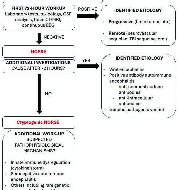
Once the diagnosis of NORSE is suspected (ie, if the initial set of examinations did not suggest a cause), further etiologic investigation must be continued without delay (Figure 1). Identification of an underlying etiology does not necessarily exclude NORSE. Autoimmune/paraneoplastic encephalitis, viral encephalitis, and selected genetic or mitochondrial disorders are recognized etiologic contexts within the NORSE spectrum (Table 1). Conversely, the identification of an acute structural, toxic, or metabolic cause of RSE is inconsistent with a diagnosis of NORSE.

Genetic testing should be considered when a mitochondrial or other genetic disorder associated with NORSE is suspected. Mitochondrial disorders have been reported as causes of NORSE. In contrast, developmental and epileptic encephalopathies (DEEs) may be associated with RSE during their disease course but represent a distinct clinical scenario and should not be equated with NORSE (Table 2). When no underlying cause is identified despite an appropriate and extensive diagnostic workup, the patient is classified as having cryptogenic NORSE (cNORSE).
Several international consensus statements have comprehensively described the investigations that should be performed in individuals with NORSE.6 Table 1 summarizes reported causes of de novo RSE and highlights clinical, biologic, EEG, imaging, and contextual features that should raise diagnostic suspicion for each condition. Table 2 summarizes the main alternative causes of new-onset status epilepticus (SE) that should prompt consideration of diagnoses other than NORSE.
Recommendations highlight that the investigational panel should be tailored to the clinical presentation, medical history, and family history, but should systematically include blood and CSF analyses covering infectious (viral and bacterial) and autoimmune panels. Metagenomic next-generation sequencing of CSF should be performed as part of the infectious workup, particularly when pleocytosis is marked and conventional pathogen testing remains negative.7 Continuous EEG monitoring is essential for both seizure management and the identification of electrographic patterns suggestive of specific pathologies, such as generalized rhythmic delta activity and extreme delta brush in anti-NMDA receptor encephalitis,8 or parieto-occipital periodic epileptiform discharges in mitochondrial disorders.9

Brain MRI
Brain MRI plays a central role in the diagnostic evaluation of NORSE. First, it may identify a structural cause of RSE, such as an acute stroke or brain tumor, thereby indicating a diagnosis other than NORSE. Second, MRI may provide clues to an underlying cause of NORSE, including autoimmune, infectious, or genetic disorders, with patterns such as mesial temporal abnormalities in autoimmune or viral encephalitis and pseudostroke in mitochondrial disease.9 A claustrum hyperintensity, meanwhile, is suggestive of cNORSE.10 Third, MRI is useful to evaluate seizure-related abnormalities, including cortical ribboning and the pulvinar sign.11 Normal MRI results do not exclude cNORSE, especially early in the disease course, and MRI should therefore be repeated when clinically indicated.12
Genetic Testing and Brain Biopsy
Genetic testing, including mitochondrial testing, and evaluation for inherited metabolic encephalopathy should be considered not only in young children, but also in adolescents and adults in whom no cause has been identified after an extensive first-line workup. Brain biopsy should be reserved for selected individuals with a targetable lesion when the diagnosis remains inconclusive.13 In cNORSE, biopsy findings remain nonspecific.
Cryptogenic NORSE
Despite an extensive workup, more than half of cases have no identified cause and are classified as cNORSE. Recent studies suggest that blood cytokine profiling may help characterize the biologic heterogeneity of these cases and identify distinct inflammatory patterns (Figure 1).14 Using an extended 96-cytokine panel, 3 cNORSE clusters have been identified: cluster A, comprising older individuals with little evidence of inflammation; cluster B, characterized by elevated innate immunity cytokines and typically associated with poor outcomes; and cluster C, characterized by dysregulated adaptive immune responses and favorable long-term recovery.14 These profiles raised hypotheses regarding differential responses to immune-targeted therapies, but their clinical utility for treatment selection has not yet been established. In particular, cytokine profiles should not be used to withhold or delay immunotherapy, and treatment should follow the 2022 international consensus recommendations.4
At our institution, cytokine profiling is used as a complementary tool, particularly when considering second-line immunotherapy, whereas treatment decisions remain primarily based on the clinical presentation. Individuals can be assigned to an inflammatory cluster using graph-based analysis,15 and a proxy 12-cytokine panel (interleukin-1β [IL-1β], interleukin-2 [IL-2], interleukin-4 [IL-4], interleukin-6 [IL-6], C-X-C motif chemokine ligand 8/interleukin-8 [CXCL8/IL-8], interleukin-10 [IL-10], interleukin-12p70 [IL-12p70], interleukin-17A [IL-17A], tumor necrosis factor-α [TNF-α], C-C motif chemokine ligand 2 [CCL2], C-C motif chemokine ligand 3 [CCL3], and granulocyte colony-stimulating factor [G-CSF]) provides a simplified approach for clinical investigation.16
Interestingly, some structural or neoplastic lesions may trigger secondary inflammatory responses that contribute to RSE. Whether such immune activation can result in a NORSE-like inflammatory phenotype remains speculative. Cytokine profiling may represent an exploratory approach to characterize these inflammatory responses, but its clinical and mechanistic relevance remains to be established.

Symptomatic Management in the ICU
The symptomatic management of NORSE builds upon the 2022 international expert recommendations for NORSE and the 2018 guidelines for SE management.4,17 Treatment intensity should be individualized according to SE severity and semiology: refractory generalized tonic-clonic SE warrants rapid therapeutic escalation, whereas refractory focal and nonconvulsive SE require a more nuanced approach based on seizure burden, EEG evolution, and clinical context, ideally in consultation with an epileptologist. This is particularly relevant in NORSE, where seizures are frequently nonconvulsive and higher seizure burden has been associated with an increased risk of post-NORSE epilepsy.18
Management of SRSE and prolonged SRSE, commonly observed in cNORSE, is complex and may extend over several weeks, exposing individuals to sedation- and ICU-related complications that, in addition to the underlying pathogenesis, represent a major cause of mortality. Therefore, identifying the optimal depth and duration of anesthesia to maximize neuroprotection while minimizing treatment-related complications is a key therapeutic objective. A structured, stepwise, and multipronged approach based on 7 principles—monitoring, antiseizure medication (ASM) optimization, anesthetic management, other therapeutic strategies, adverse effect monitoring, continuous intravenous anesthetic drug (CIVAD) weaning, and management of recurrent SE—can help guide care.19
Seven Principles to Guide Care
Monitoring
All people with superrefractory NORSE require specialized ICU admission with continuous video-EEG monitoring to enable real-time titration of CIVADs and optimization of ASMs based on seizure activity and sedation depth. This level of monitoring is also essential for identifying and monitoring patterns along the ictal–interictal continuum, whose clinical significance and contribution to ongoing brain injury remain incompletely understood.20 Close collaboration with experienced neurophysiologists, together with serial brain MRI to assess seizure-related lesions and their evolution, is essential to guide treatment decisions.
Antiseizure Medication Optimization
ASMs are systematically maintained throughout the course of SRSE despite failure of first-line treatment. We optimize the ASM regimen using a mechanism-based strategy, combining ≤3 ASMs with different mechanisms of action rather than multiple agents acting primarily on the same molecular target. This approach aims to target distinct pathways involved in seizure generation and propagation while limiting overlapping adverse effects.
ASMs with the lowest potential for drug–drug interactions are preferred. In our institution, levetiracetam (3 to 4 g/d) is used first, with the subsequent addition of lacosamide (400 mg/d), and, if needed, perampanel (12 mg/d). If SE persists or recurs, further options, including valproic acid, topiramate, cannabidiol, phenytoin, zonisamide, or phenobarbital, may be considered. All ASMs must be prescribed at therapeutically effective doses, with appropriate loading doses. Frequent monitoring of ASM serum concentrations is recommended because of the high potential for drug–drug interactions and the variability in ASM metabolism, particularly due to increased renal clearance, which is frequently observed in SRSE.21
Anesthetic Management
CIVADs are titrated to achieve clinical and electrographic seizure suppression or, when this cannot be achieved, to induce a burst-suppression pattern.19 CIVADs should initially rely on midazolam or propofol, which may be combined when either agent alone is insufficient. When seizures remain uncontrolled despite optimized propofol and midazolam therapy, several anesthetic strategies can be considered. Our institutional practice is to use ketamine (or esketamine) and inhaled volatile anesthetics (mainly isoflurane) as second-line anesthetic options. We reserve barbiturates (thiopental or pentobarbital) for the most refractory cases because of their substantial adverse effect profile, including prolonged coma, cardiovascular instability, immunosuppression, ileus, and thrombosis (Table 3). Except for barbiturates, CIVADs are often used in combination to limit dose escalation, with close monitoring for cumulative adverse effects. ASMs are systematically maintained alongside CIVADs to ensure seizure control.
Other Therapeutic Strategies
Other therapeutic strategies include a ketogenic diet (KD), which should be considered within 7 days, or earlier when a mitochondrial or other metabolic disorder potentially responsive to the KD is suspected. Specific inborn errors of metabolism, including disorders of fatty acid oxidation, primary carnitine deficiency, and pyruvate carboxylase deficiency, contraindicate the KD. Propofol should be avoided during the KD because of the increased risk of propofol infusion syndrome. Blood glucose, ketone levels, and the lipid profile are closely monitored.
Neuromodulation may also be considered in selected cases of NORSE.19 Vagus nerve stimulation represents a relatively low-risk option in NORSE.22 Acute deep brain stimulation and responsive neurostimulation have been reported, particularly after resolution of SE, but remain exceptional therapies.23,24

Adverse Effect Monitoring
Systematic monitoring of adverse effects of ASMs and CIVADs is essential and should include complete blood count, electrolytes, acid-base status, lactate, creatine kinase, triglycerides, and liver function tests. Acute colonic pseudo-obstruction (Ogilvie syndrome) is a potentially serious complication that requires careful prevention and close monitoring of bowel function. To reduce this risk, the concomitant use of opioids and high-dose sedative therapy should be avoided.
CIVAD Weaning
The background ASM regimen must be fully optimized before initiating anesthetic weaning, including an enteral benzodiazepine (ie, clobazam) administered in 3 or 4 divided doses per day. Clobazam is used because of its sustained antiseizure activity and relatively limited sedative properties compared with other benzodiazepines.25 Anesthetic agents are tapered gradually, one agent at a time, every 6 to 12 hours under continuous EEG monitoring. This approach reflects our institutional practice, as no guideline-level evidence currently supports a specific anesthetic-weaning strategy in RSE.
Management of Recurrent Status Epilepticus
During anesthetic weaning, recurrence of electrographic or clinical SE should prompt immediate reinstitution of the last effective anesthetic dose, maintained for approximately twice the duration of the preceding weaning attempt before a slower taper is reattempted under continuous EEG monitoring. In contrast, isolated seizures without recurrence of SE do not warrant prolongation or escalation of CIVAD therapy.
Immunotherapy
Immunotherapy should be initiated promptly, once major infectious causes have been reasonably excluded, without waiting for completion of an exhaustive diagnostic workup. The 2022 international recommendations advocate for initiating first-line immunotherapy within the first 72 hours and escalating to second-line agents within 7 days in cases of refractory cNORSE.4 As a tertiary referral center specializing in refractory cases, our institution receives some individuals beyond these time windows. In such cases, immunotherapy is initiated or escalated as early as possible after admission.
First-Line Immunotherapy
In our institution, we typically administer 5 initial daily boluses of high-dose methylprednisolone (1000 mg/d), with 2 to 5 additional boluses depending on clinical response and tolerance, followed by oral corticosteroids. Corticosteroids may be combined with plasma exchange, generally administered for 5 to 10 sessions over ~1 to 2 weeks using albumin-based replacement fluid, although the number and interval of exchanges may be adapted according to clinical response and tolerability. Alternatively, intravenous immunoglobulins are administered at 2 g/kg over 2 to 5 days with adequate hydration. Particular caution is warranted in individuals with cardiac or renal insufficiency or an increased thrombotic risk.
Second-Line Immunotherapy
In individuals with cNORSE who remain refractory despite first-line treatment, second-line treatments should preferably be initiated within 7 days, in accordance with the 2022 consensus recommendations.4 When antibody-associated autoimmune encephalitis is diagnosed or strongly suspected based on established clinical criteria and autoantibody testing,26 treatment is tailored to the underlying disease. Rituximab is generally preferred for encephalitis associated with neuronal surface antibodies, whereas cyclophosphamide may be considered particularly in diseases associated with intracellular antigens. In seronegative cases, the diagnosis of probable autoimmune encephalitis should rely on established clinical criteria rather than cytokine profiling.
When neither antibody-associated autoimmune encephalitis nor a mitochondrial disorder is suspected, the 2022 consensus recommendations support the use of cytokine-targeted therapies, particularly anakinra (Kineret; Swedish Orphan Biovitrum AB, Waltham, MA) or tocilizumab, in cNORSE.4 At our institution, we preferentially use anakinra (anti–IL-1) at 10 mg/kg/d intravenously (IV) or subcutaneously (SC) (maximum 400 mg/d). If there is no clinical response after 2 to 3 weeks, or if anakinra is contraindicated, tocilizumab (anti–IL-6) may be considered at 8 to 12 mg/kg IV every 2 weeks. The combination of anakinra and tocilizumab is avoided to limit the cumulative risk of serious infections.
Intrathecal corticosteroids (dexamethasone or methylprednisolone) represent another therapeutic option in refractory NORSE and have also been used in antibody-mediated autoimmune encephalitis.27,28 At our institution, 4 to 8 administrations are typically given every 2 days. Their limited systemic immunosuppressive effect may make them particularly relevant when systemic immunosuppression is undesirable, including in individuals with concurrent infection or cytopenia. These agents may be used alone or in combination with systemic immunotherapy according to the clinical context.
Cytokine Profiling
Cytokine profiling is not established as a validated tool for selecting immunotherapy and should not replace clinical assessment, autoantibody testing, or consensus recommendations. At our institution, a 12-cytokine panel is performed systematically and longitudinally in serum and CSF as an investigational approach to characterize the inflammatory response and potentially inform treatment decisions, particularly when considering therapies targeting innate immune pathways. Preliminary studies suggest that distinct inflammatory profiles may identify biologically different subgroups of cNORSE, but whether cytokine-guided treatment improves outcomes remains unknown and requires prospective validation.14,15
After ICU Discharge
Although cNORSE is generally a monophasic acute illness, its consequences frequently persist long after SE has resolved. Approximately 20% of individuals will die during the acute phase, with mortality rates being higher in adults than in children.29 Among survivors, sequelae of the acute brain injury commonly include post-NORSE epilepsy, cognitive and functional disabilities, and reduced health-related quality of life.30 These outcomes highlight the need for structured postacute care, including epilepsy management, neurologic and cognitive follow-up, and multidisciplinary rehabilitation.
ASM Reduction Strategy
As soon as SE is resolved and CIVADs are weaned, individuals should, whenever feasible, be managed in a center with expertise in epilepsy. ASM reduction should be individualized according to the severity of the acute illness, the electrographic pattern, and the occurrence of recurrent seizures. Guided by clinical and EEG findings, ASMs can be gradually reduced over weeks, starting preferentially with the most sedating drugs, including benzodiazepines. Further tapering may continue after hospital discharge over several months.
Approximately 70% of individuals with cNORSE develop post-NORSE epilepsy, which is frequently drug-resistant.30 Therefore, the goal is not necessarily to achieve complete seizure freedom at the cost of excessive polytherapy, but rather to balance seizure control with treatment-related adverse effects. EEG monitoring should be repeated when clinically indicated, particularly when altered behavior or cognition raises concern for recurrent nonconvulsive seizures.
Evaluation of the Consequences of NORSE
Once SE has resolved and the individual is clinically improving, management shifts from the acute investigation and treatment of NORSE toward assessment of its neurologic consequences. At this stage, the clinical picture may reflect a combination of sequelae from the acute brain injury, the emergence of a potentially epileptogenic network, and the effects of prolonged intensive care and ASM exposure. Individuals with persistent or progressive neurologic abnormalities suggesting ongoing disease activity may require renewed investigations into the underlying pathology, including repeat malignancy screening or further autoimmune/paraneoplastic, infectious, or genetic testing, as appropriate.
In individuals recovering from the acute phase, neurologic follow-up should include detailed cognitive assessment, with particular attention to memory and executive functions. Brain MRI may show residual or evolving abnormalities related to the acute brain injury. Bilateral hippocampal signal abnormalities and swelling observed during the acute phase may evolve toward hippocampal atrophy, which is associated with severe memory impairment and a high subsequent seizure burden.12,31 After prolonged cNORSE, diffuse cortical and subcortical atrophy may represent chronic findings and is associated with more extensive cognitive and functional disabilities.32 In individuals with autoimmune encephalitis, inflammation likely lasts longer than in individuals with cNORSE, and inflammatory activity can evolve within lesions that appear chronic. Further investigation or treatment should be considered when the clinical, biologic, or imaging course raises concern for an ongoing disorder, such as autoimmune encephalitis or a paraneoplastic syndrome.
Psychiatric and behavioral complications are frequent and may result from post–intensive care syndrome, ASM adverse effects, direct brain injury, or psychologic reactions to severe neurologic deficits, particularly when familial or professional functioning is affected.33
Neurologic Rehabilitation
Individuals with disabilities should be referred to neurologic rehabilitation after the ICU stay for intensive rehabilitation, including neuropsychology, speech therapy, physiotherapy, and occupational therapy. Recovery after cNORSE is highly variable and may continue over months to years. Although many survivors experience persistent cognitive and functional impairments and reduced quality of life, substantial recovery is possible, with some individuals regaining independence and returning to education or employment.30,34
Specific Therapies
In individuals in whom autoimmune encephalitis was diagnosed, immunosuppressive drugs should be continued according to practice recommendations, which depend on the subtype of autoantibodies, and screening for cancer must be performed in the context of paraneoplastic autoimmune encephalitis.35
When immunotherapy is considered to have been effective during the acute phase, the 2022 international consensus recommendations support continuing immunomodulatory treatment into the postacute phase for at least 3 months.4 Treatment can subsequently be tapered according to the clinical course and tolerability. If neurologic or seizure-related symptoms significantly worsen during or after immunotherapy withdrawal, resumption of the previously effective treatment should be considered.4 However, the optimal duration of immunotherapy after NORSE remains unknown, and some individuals may require longer treatment, particularly when there is evidence of persistent or relapsing immune-mediated disease.
At our institution, longitudinal serum and CSF cytokine profiling is performed as an investigational tool to characterize the evolution of the inflammatory response after cNORSE. Although normalization of cytokine abnormalities may provide information on biologic changes over time,36 it should not be interpreted as evidence that immunotherapy is no longer beneficial or used in isolation to determine treatment duration. The relationship among persistent inflammation, post-NORSE epilepsy, and response to prolonged immunotherapy remains incompletely understood, and prospective studies are needed to determine whether biomarkers can help individualize treatment duration.
Conclusion
NORSE is a rare, devastating condition requiring rapid identification, aggressive acute management, and structured postacute rehabilitation. Coordinated multidisciplinary care, standardized recommendations, and dedicated reference centers are essential to improve long-term neurologic and functional outcomes.
Ready to Claim Your Credits?
You have attempts to pass this post-test. Take your time and review carefully before submitting.
Good luck!
