
Electrodiagnostic Testing for Cervical and Lumbosacral Radiculopathy
Recognizing common pitfalls in EMG and nerve conduction studies can improve diagnostic accuracy and help avoid false-positive and false-negative findings in cervical and lumbosacral radiculopathy.
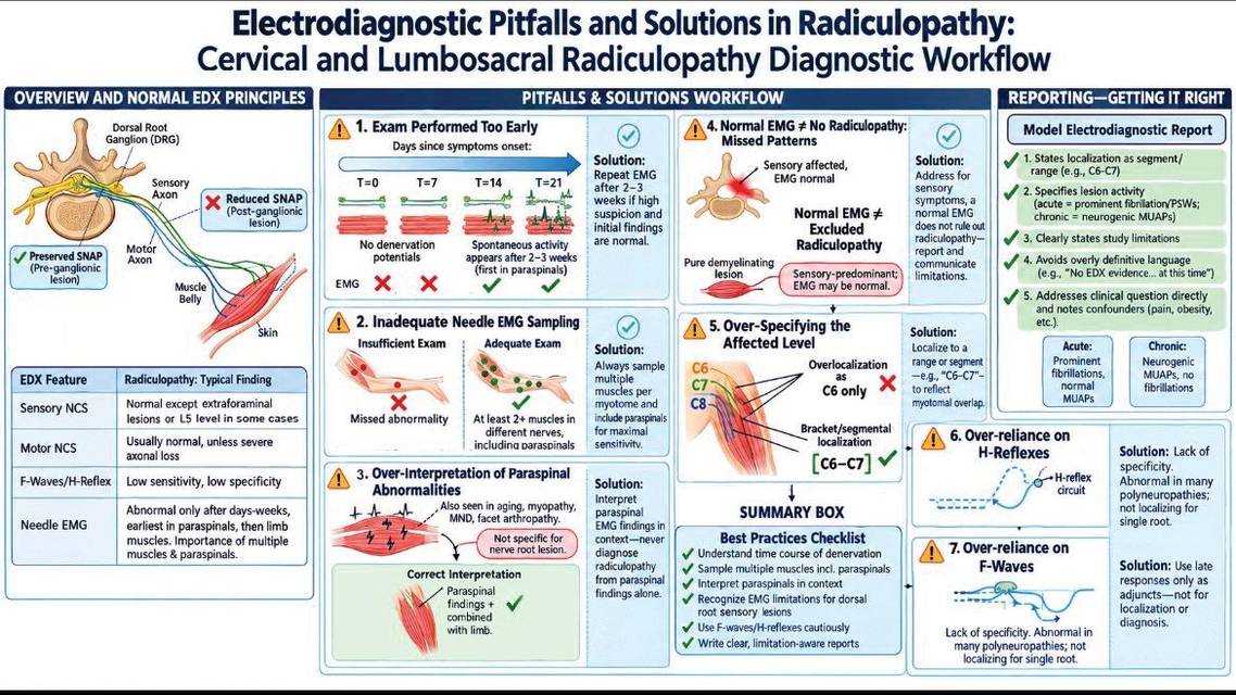
KEY TAKEAWAYS
- Timing can significantly affect electrodiagnostic testing for radiculopathy, and an early normal EMG does not necessarily exclude clinically meaningful nerve root disease.
- Muscle selection, paraspinal findings, myotomal overlap, and postsurgical changes can all create diagnostic pitfalls when EMG and nerve conduction study results are interpreted too narrowly.
- High-quality electrodiagnostic reporting should define lesion activity and chronicity while acknowledging test limitations and integrating findings with the neurologic examination and imaging.
Common Pitfalls and Solutions
Cervical and lumbosacral radiculopathies are among the most common conditions referred for electrodiagnostic evaluation, and the accurate interpretation of these studies is paramount for optimal patient care. Electrodiagnostic studies serve a key role in objectively confirming a nerve root lesion, excluding differential diagnoses, and providing prognostic information through assessment of activity, chronicity, and severity of the lesion.
The sensitivity of electrodiagnostic studies has been reported as 50% to 71% for cervical radiculopathy and 49% to 86% for lumbosacral radiculopathy, with specificity of 87% to 100% when appropriate diagnostic criteria are used.1 This wide range reflects not only limitations of the studies themselves but also the potential for misinterpretation. Even experienced electrodiagnostic consultants can encounter subtle, incomplete, or potentially misleading findings. Errors generally arise from 2 interrelated sources: errors in the technical execution and interpretation of the findings and errors in reporting that may convey an inaccurate or misleading impression to the referring physician. These interpretive lapses carry serious clinical consequences, including false-negative or false-positive diagnoses, unnecessary surgeries, delayed recognition of alternative neurologic disorders, and inappropriate patient management.
Over the past several decades, numerous studies have evaluated the diagnostic performance of individual electrodiagnostic techniques. However, relatively few reviews have focused specifically on the practical diagnostic errors encountered during electrodiagnostic evaluation and the strategies that can minimize them in daily clinical practice. This review aims to guide the clinician through the most common pitfalls inherent in the electrodiagnostic evaluation of radiculopathy, providing a framework for avoiding these errors and enhancing the diagnostic accuracy and clinical utility of the electrodiagnostic report.
Basic Electrodiagnostic Principles in Radiculopathy
The location of the dorsal root ganglion (DRG) has important electrodiagnostic implications. In most nerve roots, the DRG lies within or just distal to the intervertebral foramen; therefore, compressive root lesions typically occur proximal to the DRG.2,3 Because the sensory neuron cell body and distal axon remain intact, distal sensory nerve conduction studies (NCS) are usually preserved despite sensory symptoms. This preservation of sensory nerve action potentials (SNAPs) distinguishes radiculopathy from postganglionic lesions, such as plexopathy or entrapment neuropathy, in which SNAPs are reduced or absent.2-4 Exceptions exist, notably L5 root lesions, in which the superficial peroneal SNAP may be affected due to an intraforaminal DRG location in the L5 level,5,6 as well as some far-lateral and extraforaminal lesions.7
Motor NCS are also usually normal, as root lesions often affect only a subset of motor axons and compensatory innervation preserves compound muscle action potential (CMAP) amplitude.2-4 However, severe axonal loss may reduce CMAP amplitudes in affected muscles. Conduction velocities and distal latencies remain normal because lesions are primarily axonal.8 Late responses have limited sensitivity. F-waves are abnormal only in severe lesions or those involving multiple roots and lack localizing value. The soleus H-reflex may support S1 radiculopathy but lacks specificity.2-4,8 The primary utility of NCS in radiculopathy is therefore excluding alternative diagnoses, such as entrapment neuropathies, plexopathies, and peripheral neuropathies.
Needle EMG is the cornerstone of electrodiagnostic confirmation, providing direct evidence of motor axonal injury.2,3 Needle EMG evaluates 3 key measures: spontaneous activity, motor unit action potential morphology, and recruitment pattern.
 Spontaneous Activity
Fibrillation potentials and positive sharp waves (PSWs) indicate active axonal loss and are the earliest reliable signs, appearing in paraspinal muscles within the first week and limb muscles by 2 to 3 weeks.2,3,8
Motor Unit Action Potential Morphology
Persistent denervation with collateral reinnervation produces motor unit action potentials (MUAPs) that are increased in amplitude, prolonged in duration, and polyphasic, signifying neurogenic remodeling over weeks to months.2-4
Recruitment Pattern
Reduced MUAP recruitment in weak muscles reflects loss of functional motor axons and is particularly important in the earliest phase of injury, before spontaneous activity develops.2-4,8
Diagnosis requires abnormalities in at least 2 muscles innervated by the same root but different peripheral nerves, with normal findings above and below the suspected level.9,10 This confirms the abnormality is localized to the nerve root rather than a single peripheral nerve or plexus.
Paraspinal muscles deserve attention because they receive direct innervation from the dorsal primary rami shortly after the spinal nerve exits the foramen. Denervation may appear there earlier than in distal limb muscles. However, paraspinal abnormalities are neither universally present nor entirely specific, as age-related changes, previous surgery, technical factors, and longstanding degeneration may reduce specificity.2-4,8
Common Electrodiagnostic Pitfalls and Practical Solutions
Common electrodiagnostic pitfalls and practical approaches to avoiding them are summarized in the Table.
Performing the Examination Too Early After Symptom Onset
A major cause of false-negative electrodiagnostic examinations is failure to recognize the temporal evolution of axonal degeneration and muscle denervation. Immediately following acute root injury, conduction block and impaired axonal transport may produce pain, weakness, or sensory symptoms without detectable spontaneous activity on EMG or a drop in CMAP amplitude on NCS. Spontaneous activity on EMG—namely, fibrillation potentials and PSWs—appears in a proximal-to-distal sequence, with abnormalities detectable in paraspinal muscles within the first week, whereas spontaneous activity becomes detectable in limb muscles after ~2 to 3 weeks. During the first 10 to 14 days, distal motor axons remain intact, and CMAP amplitudes are unaffected because Wallerian degeneration has not yet reached the muscle.2-4,11 Consequently, in the earliest phase of nerve root lesions, the only electrodiagnostic finding may be reduced MUAP recruitment in weak muscles, reflecting the loss of functional motor axons.
A normal or equivocal EMG result obtained within the first 1 to 2 weeks of symptom onset should be interpreted with caution; it does not exclude radiculopathy and may simply reflect insufficient time for spontaneous activities to develop. When clinical suspicion remains high, repeat examination after an appropriate interval often reveals abnormalities that were initially absent.
Performing an Inadequate Needle EMG Examination
A key technical source of false-negative electrodiagnostic studies is the failure to sample an adequate number of muscles within the affected myotome. This pitfall arises from the segmental innervation pattern. A single spinal nerve root contributes to multiple muscles through several peripheral nerves, and a nerve root lesion may preferentially affect fascicles supplying some muscles while leaving others intact.12 Consequently, EMG findings may be inconsistent across muscles of the same myotome, with EMG abnormalities evident in some and not in others. Sampling a greater number of muscles per myotome enhances diagnostic sensitivity13,14; however, the electromyographer must balance this against patient discomfort and the practical constraints of examination time. Paraspinal muscle sampling is also an important component of the examination, particularly when clinical suspicion remains high despite normal findings in limb muscles on EMG, as a substantial proportion of radiculopathies may be missed if paraspinal muscles are not included in the electrodiagnostic evaluation of suspected root disease.15-17
Misinterpreting Paraspinal Muscle Findings
Paraspinal muscle examination is a valuable component of the needle EMG evaluation of radiculopathy, but paraspinal EMG is frequently overinterpreted. First, spontaneous activity confined to paraspinal muscles does not necessarily indicate radiculopathy. Sparse fibrillation potentials and PSWs lasting <3 seconds may be present in the paraspinal muscles of asymptomatic individuals, particularly in people >40 years of age.18,19 Recognizing this age-related phenomenon is essential to avoid overdiagnosing radiculopathy based on such nonspecific findings. Furthermore, fibrillation potentials and PSWs in the paraspinal muscles are not always a result of nerve root–level lesions. They can occur in other conditions, such as proximal myopathies, motor neuron disorders, and even facet joint arthritis with dorsal ramus entrapment.20-22
Moreover, normal paraspinal EMG findings do not always exclude radiculopathy. Selective dorsal root involvement, longstanding reinnervation, obesity limiting needle placement, or inadequate sampling may all reduce the sensitivity of paraspinal examination.
The diagnostic value of paraspinal EMG is greatest when abnormalities occur together with neurogenic findings in appropriately selected limb muscles belonging to the same myotome. Paraspinal EMG abnormalities should therefore be interpreted cautiously and should not, in isolation, be considered sufficient evidence of radiculopathy.
Assuming That Normal Needle EMG Results Exclude Radiculopathy
A common misconception is that a normal EMG result rules out cervical or lumbosacral radiculopathy. Several clinical situations, including isolated dorsal root lesions and purely demyelinating lesions, may produce normal EMG results despite existing nerve root pathology.
Isolated Dorsal Root Lesions. Radicular pain may arise from isolated compression of dorsal root sensory fibers, leaving ventral motor axons unaffected. In this setting, EMG, which primarily assesses motor unit function, may be normal despite substantial symptoms. This mismatch between clinical severity and electrodiagnostic normality constitutes a common pitfall, potentially leading to false-negative results and delayed diagnosis. Clinicians should therefore be aware that normal electrodiagnostic study results do not rule out a sensory-predominant compressive radiculopathy.23
Purely Demyelinating Lesions. When nerve root compression produces demyelination and conduction block in the absence of axonal injury, EMG will show no spontaneous denervation potentials. Reduced recruitment of MUAPs in clinically weak myotomal muscles may be the only electrodiagnostic abnormality.24,25 This pattern, frequently encountered in the early phase of compressive radiculopathy, carries a risk of misinterpretation as a false-negative study. It thus exemplifies an important constraint of EMG in the evaluation of demyelinating root lesions.
Clinicians must recognize the inherent limitations of electrodiagnostic studies in detecting root-level pathology, particularly when clinical suspicion remains high. Normal electrodiagnostic study results in an individual with convincing radicular symptoms do not indicate a flawed examination; rather, they indicate that electrodiagnostic abnormalities are not detectable at that particular moment. Clear acknowledgment of this limitation in the final report not only strengthens the clinician's credibility but also promotes effective communication with the referring provider. A suitable reporting phrase would be "there is no electrodiagnostic evidence of an active or chronic nerve root lesion on this study," accompanied by a note that this finding does not exclude a clinically significant radiculopathy.

Overrelying on EMG for Precise Single-Level Localization
A common and potentially harmful pitfall in electrodiagnostic practice is the tendency to overspecify the exact spinal root level of a lesion based on needle EMG findings alone. Whereas EMG can confirm the presence of radiculopathy, its precision for single-level localization is limited by anatomic realities. Myotomal anatomy exhibits considerable overlap and interindividual variability. Most limb muscles receive contributions from multiple spinal roots. For instance, the triceps brachii is primarily innervated by C7 but often receives substantial C6 and C8 contributions. Therefore, abnormalities in a single muscle cannot reliably identify the exact affected nerve root. The overlapping nature of myotomal innervation means that EMG findings typically implicate a spinal segment (eg, C6–C7 or L5–S1) rather than an isolated, single root.9,13,14,24
To address this limitation, practitioners should sample myotomes above and below the suspected level to bracket the abnormality and confirm focal pathology. Accurate reporting should frame findings as involving a spinal segment (eg, a “C6–C7 nerve root lesion”) rather than using overspecified localizations (eg, an “isolated C6 nerve root lesion”), thereby acknowledging the overlapping nature of myotomal innervation and the limited precision of EMG.
Electrodiagnostic findings should be integrated with clinical and imaging data, with EMG serving to confirm the neurophysiologic significance of MRI abnormalities. The electrodiagnostic consultant should not claim a level of precision that the test cannot deliver, and if EMG localizes the lesion to a segment whereas imaging shows single-level pathology, the report should acknowledge this synergy and the complementary nature of the studies.
Overrelying on F Waves and H Reflexes
Late responses have traditionally been included in the electrodiagnostic evaluation of radiculopathy because they assess conduction along proximal motor pathways. However, their diagnostic limitations are frequently underestimated.
F-wave studies have low sensitivity for radiculopathy because the response represents the summed conduction of multiple motor axons originating from several spinal roots. A single-root lesion often fails to produce measurable abnormalities because most muscles receive innervation from multiple adjacent nerve roots. Moreover, F-wave abnormalities lack anatomic specificity because the F wave traverses a long pathway from the muscle to the peripheral nerve, nerve root, and spinal segment. F-wave abnormalities may occur in peripheral neuropathies, plexopathies, or generalized motor neuron disorders. Therefore, abnormal F-wave findings should be regarded as supportive rather than diagnostic evidence.26,27
The soleus H reflex has greater clinical utility, particularly in suspected S1 radiculopathy. Delayed or absent H reflexes may provide objective evidence of S1 root dysfunction even when routine NCS results remain normal. Nevertheless, the H reflex is influenced by age, limb length, peripheral neuropathy, and technical factors. Therefore, bilateral absence of H reflexes in an older individual should not automatically be interpreted as bilateral S1 radiculopathy.28
Electrodiagnostic Challenges in the Postsurgical Population
Interpretation of electrodiagnostic studies becomes considerably more complex in individuals with previous spinal surgery. After spinal surgery, persistent denervation changes (high-amplitude, long-duration, and polyphasic MUAPs) may persist indefinitely despite successful nerve root decompression.29,30 Therefore, longstanding neurogenic MUAP changes alone should not be interpreted as evidence of recurrent radiculopathy. Instead, active denervation or newly developed abnormalities in previously unaffected muscles are more suggestive of ongoing or recurrent nerve root injury.
Paraspinal muscle examination deserves special consideration in the postsurgical population. Surgical dissection often results in denervation of the posterior paraspinal muscles, producing abnormal spontaneous activity on EMG that is unrelated to ongoing nerve root compression. Such abnormalities may persist for years after spinal surgery, even in individuals without clinical symptoms.31,32 As a result, whereas the presence of PSWs and fibrillation potentials in the paraspinal muscles is a highly localizing finding in individuals without previous surgery, its diagnostic reliability is diminished in the postsurgical population because of iatrogenic muscle injury and scar tissue.
To avoid this pitfall, in the postsurgical population, diagnostic emphasis must shift to the limb muscles, where active denervation constitutes the most compelling evidence of recurrent or ongoing nerve root lesions. Longstanding neurogenic MUAP changes in limb muscles may simply reflect the original presurgical lesion rather than recurrence. The key finding supporting a true recurrent or ongoing root lesion is the coexistence of new, active denervation, manifesting as high-amplitude fibrillation potentials and PSWs, alongside these longstanding changes, indicating an ongoing, active nerve root injury.
Pitfalls in Generating Electrodiagnostic Reporting
Even when the electrodiagnostic examination is technically well-performed and correctly localized, its clinical utility can be substantially undermined by reporting errors, incomplete documentation, or ambiguous conclusions. These deficiencies may render a study less useful for clinical decision-making or, worse, mislead the referring physician. Several common reporting pitfalls warrant particular attention.
Overstating the Pathogenesis of the Lesion
A frequent reporting error is the attribution of electrodiagnostic findings to a specific anatomic or structural cause. Stating an impression such as “electrodiagnostic evidence of a chronic right C6 disc herniation” oversteps the capabilities of the electrodiagnostic study. The electrodiagnostic study characterizes the neurophysiologic state of the nerve root, not its underlying structural pathogenesis. A nerve root lesion may result from various pathologies, including facet joint hypertrophy, spondylolisthesis, inflammatory or infectious processes, synovial facet cysts, or neoplastic lesions.33
The impression should therefore accurately reflect the neurophysiologic diagnosis. The electrodiagnostic consultant should use precise, neurophysiologic terminology, such as “electrodiagnostic evidence of a chronic right C6 nerve root lesion.” The specific cause of the lesion cannot be determined by electrodiagnostic study results alone and requires correlation with clinical history and imaging findings.
Incompletely Characterizing the Lesion
A report that merely identifies the presence of a nerve root lesion without elaborating on its characteristics represents a lost opportunity to inform patient management. A statement such as "this study shows right C6 radiculopathy" offers little more than a binary diagnosis and does not address the activity, chronicity, or severity of the underlying pathology.
The electrodiagnostic report should provide a descriptive interpretation of activity and chronicity based on objective findings. Activity (ie, active axonal loss) can be determined by the presence and amplitude of fibrillation potentials and PSWs. Higher amplitudes are typically associated with recent or ongoing axonal injury.2,3,22 Chronicity can be determined by the presence of persistent neurogenic MUAP changes, including increased amplitude, prolonged duration, and increased polyphasia.
The characterization of activity and chronicity hinges on the dynamic interplay between abnormal spontaneous activity and MUAP morphology. In an acute lesion, high-amplitude fibrillation potentials and PSWs reflect recent denervation. Over time, as collateral reinnervation occurs, the amplitude of these spontaneous potentials diminishes. Concurrently, MUAPs remodel into large, polyphasic, and long-duration units, signifying chronicity.1,2,4 This progression allows for recognition of 3 primary clinical scenarios: acute lesions, characterized by high-amplitude fibrillation potentials and PSWs without substantial longstanding MUAP changes, indicating acute axonal loss; chronic stable lesions, characterized by low-amplitude fibrillation potentials and PSWs with longstanding neurogenic MUAP remodeling, suggesting a previous injury that is no longer active; and acute-on-chronic or ongoing lesions, characterized by the coexistence of both high- and low-amplitude spontaneous activity alongside persistent neurogenic MUAP changes, indicating a previously injured nerve root undergoing new or ongoing axonal injury—a key distinction that signifies a new injury superimposed on an old lesion.
Reports that clearly characterize lesions as acute, chronic, or ongoing provide actionable information for surgical and rehabilitative planning, thereby enhancing the clinical utility of the electrodiagnostic consultation.
Overstating Diagnostic Certainty and Underemphasizing Clinical Relevance
Overly definitive language represents another pitfall. Because the sensitivity of needle EMG is imperfect, statements such as “radiculopathy is excluded” should generally be avoided. A more accurate conclusion is “there is no electrodiagnostic evidence of axonal nerve root lesion at the time of examination.” This wording acknowledges the limitations of the study while providing clinically meaningful guidance.
Failure to discuss study limitations may also lead to misunderstanding. Reports should indicate when interpretation is affected by factors, such as severe pain limiting muscle activation, previous surgery, obesity preventing adequate paraspinal examination, coexisting generalized peripheral neuropathy, or inability to examine key muscles.
Electrodiagnostic reports should answer the referring physician’s clinical question whenever possible. Rather than simply listing abnormal results, the conclusion should address localization, chronicity, evidence of active denervation, differential diagnosis, and important alternative explanations for the symptoms.
A high-quality report is not merely a record of electrical findings—it is a clinical consultation that integrates anatomy, physiology, and patient presentation into a coherent diagnostic assessment.
Conclusion
The electrodiagnostic examination is an extension of the clinical evaluation for radiculopathy. Its value lies not in providing a simple binary answer but in offering a detailed physiologic profile of the nerve root. Awareness of timing limitations, careful muscle sampling strategies, disciplined terminology, and correlation with clinical and imaging data are essential for accuracy. By adhering to a structured diagnostic framework, electrodiagnostic consultants can avoid misdiagnosis and deliver nuanced, clinically actionable reports that enhance patient care.
Ready to Claim Your Credits?
You have attempts to pass this post-test. Take your time and review carefully before submitting.
Good luck!
Recommended
 Epilepsy & Seizures
GAD65-Associated Temporal Lobe Epilepsy: Diagnosis and Management
Julien Hébert, MD, MSc, FRCPCJulien Hébert, MD, MSc, FRCPC