
Media formats available:

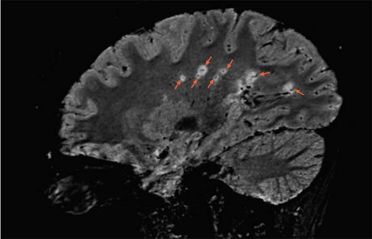
A sagittal 3T FLAIR* brain MRI shows multiple lesions with the central vein sign (orange arrows), appearing as hypointense dots or lines, located centrally within the lesion, depending on the blood vessel orientation. The FLAIR* technique was developed by Daniel S. Reich, MD, PhD and colleagues as described in Sati P, et al. Radiology. 2012;265(3):926-932. Image contributed by Daniel S. Reich, MD, PhD.
Completing the pre-test is required to access this content.
Completing the pre-survey is required to view this content.
Ready to Claim Your Credits?
You have attempts to pass this post-test. Take your time and review carefully before submitting.
Good luck!
Recommended
 Imaging & Testing
Neuroimaging: Clinical Impact of Imaging Findings
Laszlo Mechtler, MD, FAAN, FEAN, FASN, FAHSLaszlo Mechtler, MD, FAAN, FEAN, FASN, FAHS- Imaging & Testing
Neuroimaging After Brain Radiotherapy and Radiosurgery
Venkatesh Shankar Madhugiri, MBBS, MCh, FACS; Neil D. Almeida, MDVenkatesh Shankar Madhugiri, MBBS, MCh, FACS; Neil D. Almeida, MD