
Establishing a Sustainable Amyloid-Targeting Treatment Program: Lessons from One Institution
Providing disease-modifying therapies for individuals diagnosed with early Alzheimer disease requires careful patient selection, intensive safety monitoring
protocols, and the establishment of multidisciplinary workflows.
KEY TAKEAWAYS
- Safe delivery of lecanemab and donanemab requires multidisciplinary workflows and standardized monitoring.
- Patient selection, infusion logistics, and ARIA surveillance are central to amyloid-targeting treatment programs.
- Real-world implementation can improve access, efficiency, and safety in Alzheimer disease care.
Two amyloid-targeting therapies (ATTs) have been approved by the US Food and Drug Administration (FDA) for the treatment of mild cognitive impairment and mild dementia due to Alzheimer disease (AD): Leqembi (lecanemab; Eisai, Nutley, NJ) in 2023 and Kisunla (donanemab; Eli Lilly, Indianapolis, IN) in 2024. Both medications have shown efficacy in slowing disease progression (Table 1).1,2 The potential serious risk of amyloid-related imaging abnormalities (ARIA) initially made their clinical use controversial.
As outlined in “Building an infrastructure to administer amyloid-targeting therapies,”3 ATTs require an established infrastructure and interdisciplinary team to ensure appropriate diagnostic, treatment, and monitoring protocols can be maintained.
Real-world experience with clinical care introduces variability not seen in clinical trials. This article provides direct answers to common clinical and operational questions and highlights lessons learned from early adopters.

Real-World Experience
Patient Selection
Use of ATTs first requires identification of appropriate individuals early in the clinical course of AD (Figure 1). The clinical history should indicate clear deficiencies in short-term memory but independence in instrumental activities of daily living (eg, driving, working, functioning in the community). A Study to Confirm Safety and Efficacy of Lecanemab in Participants With Early Alzheimer’s Disease (Clarity AD, NCT03887455) and A Study of Donanemab (LY3002813) in Participants With Early Alzheimer’s Disease (Trailblazer-ALZ 2, NCT03367403) used the Mini-Mental State Examination (MMSE) to measure mild impairment. Clarity AD enrolled participants with MMSE scores ≥22/30 and Trailblazer-ALZ 2 included participants with MMSE scores ≥20/30. An alternative cognitive screening test, such as the St. Louis University Mental Status Examination (SLUMS) or Montreal Cognitive Assessment (MoCA), can be used to identify cognitive impairment. A SLUMS score of 21–25/30 or a MoCA score of 18–25/30 is typically considered to indicate mild impairment.
Appropriate medical testing should be completed to confirm that there is no metabolic cause of cognitive impairment. Then, biomarker testing for amyloid should be completed. The gold standard in clinical diagnosis remains amyloid PET. Cerebrospinal fluid (CSF) biomarkers represent an alternative evaluation method, with the ratio of analytes Aβ42/Aβ40, p-tau181/Aβ42, or t-tau/Aβ42 most strongly associated with amyloid pathology.4 Several blood-based biomarkers are also available, with tests including p-tau217 demonstrating superior accuracy for amyloid pathology. Although these tests are less invasive and may be more cost-effective than other screening methods, their accuracy is variable and may be affected by medical comorbidities, often requiring follow-up confirmation with an amyloid-PET or CSF study.5 Several insurance plans also require amyloid-PET or CSF biomarkers for authorization. ATTs are amyloid immunotherapies that target and remove β-amyloid plaque from the brain; therefore, the presence of amyloid is imperative to confirm an individual’s eligibility for ATT.
Participants excluded from clinical trials of ATT due to safety risks included those with inability to obtain an MRI scan; radiographic evidence of cerebral amyloid angiopathy (>4 microhemorrhages, 1 macrohemorrhage >10 mm, or superficial siderosis); severe white matter disease (Fazekas score of 3); stroke, transient ischemic attack, or seizure in the preceding 12 months; stroke involving a major vascular territory or >2 lacunar infarcts; history of an immunologic disorder requiring treatment with immunosuppressants, immunoglobulins, or monoclonal antibodies; an unstable medical condition that may affect or be affected by ATT; and chronic anticoagulant use.4,7 These are general recommendations for consideration, but some centers may elect to approve ATT therapy for selected individuals with these characteristics. For example, patients receiving another monoclonal antibody for a stable comorbid condition may still be considered for ATT. Some centers are prescribing ATT for patients with concomitant anticoagulant use though published appropriate use guidelines caution against the use of anticoagulants in patients receiving ATTs.

Figure 1. Diagnostic pathway to confirm eligibility for amyloid-targeting therapies (ATTs).
Abbreviations: ARIA, amyloid-related imaging abnormalities; CAA, cerebral amyloid angiopathy; CSF, cerebrospinal fluid; MMSE, Mini-Mental State Examination; MoCA, Montreal Cognitive Assessment; PET, positron emission tomography; SLUMS, St. Louis University Mental Status Examination.
Counseling Patients on the Use of ATT
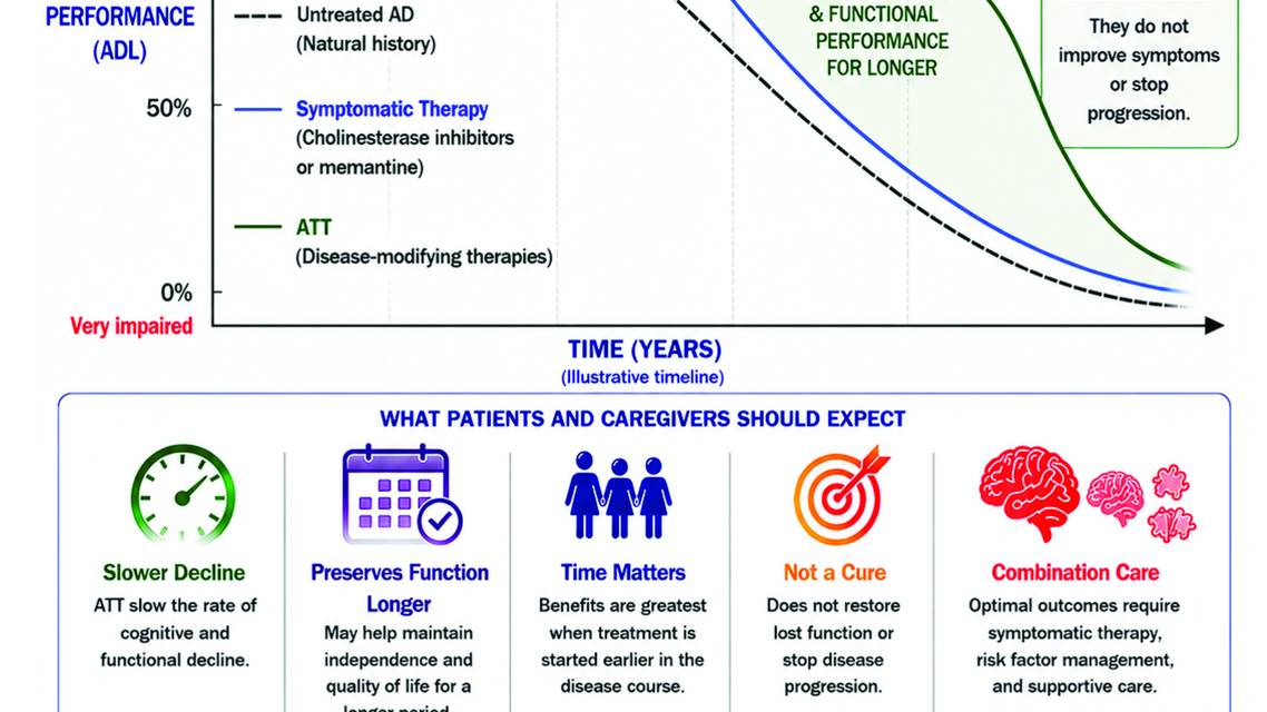
When counseling patients on the use of ATT, it is important to set realistic expectations. ATTs are not curative therapies for AD, but they have the potential to slow progression of disease (Figure 2). In Clarity AD, lecanemab demonstrated a 27% slowing of disease progression compared with placebo.1 In Trailblazer-ALZ 2, donanemab demonstrated up to 35% slowing of disease progression.2 Clinical language should highlight that slowing progression means maintaining the current level of disease (ie, mild) for a longer time, which translates to maintaining the current level of function and independence. Clinicians need to help patients and caregivers understand that ATT success is defined as slowing decline or maintaining current cognitive performance rather than improvement. In most clinical settings, routine follow-up will include repeat MMSE testing and standardized functional assessments, such as the Functional Assessment Questionnaire. These assessment tools can provide information on overall disease severity and progression.
Patients and caregivers must be educated about the risk of ARIA. ARIA is identified and monitored using surveillance MRI performed at regularly scheduled intervals to guide management (see ARIA Management). Careful patient selection using exclusion criteria (eg, radiographic evidence of severe white matter disease or cerebral amyloid angiopathy) may reduce the likelihood of treating individuals at very high risk. APOE genotype testing can also help identify individuals at high risk for ARIA, allowing clinicians to counsel patients on their personal risk.8
Initiation of ATT is based on shared decision-making among the prescribing clinician, the patient, and their caregiver. ATTs involve a complex diagnostic evaluation and treatment schedule, so an involved, well-informed caregiver is a necessity. Patients and caregivers need to understand the complexity of the treatment course. ATT requires infusion therapy 1 or 2 times per month, depending on the selected therapy, at an infusion center, and at least 4 MRI scans in the first 6 months on ATT.

Figure 2. Conceptual model of the Alzheimer disease continuum and the effects of symptomatic therapy and ATT. The untreated disease course (gray dashed line) depicts progressive cognitive and functional decline over time. Symptomatic therapies (blue line), including cholinesterase inhibitors and memantine, may provide temporary symptomatic benefit but do not alter disease progression. ATTs (green line), including lecanemab and donanemab, slow the rate of clinical decline and preserve cognitive and functional abilities for longer, but do not restore lost function or stop disease progression. The figure also highlights treatment expectations across the Alzheimer disease continuum and the importance of early diagnosis, biomarker confirmation, and timely treatment initiation.
Abbreviations: AD, Alzheimer disease; ADL, activities of daily living; ATT, amyloid-targeting therapy; MCI, mild cognitive impairment.
This figure was developed using ChatGPT 5.5 (OpenAI, San Francisco, CA).
Treatment Workflow
A standardized protocol is key to development of a feasible workflow. Before the FDA approval of lecanemab in 2023, the Norton Neuroscience Institute Memory Center (NNI) established a workgroup to build a sustainable patient care pathway for ATT. This workgroup established a care pathway (Figure 3) from initial diagnosis to starting an ATT to next steps in case of ARIA development, which can be modified and updated as needed. Memory centers may use different models to address their unique patient populations and workflows.
Use of the electronic medical record (EMR) is essential. As such, we have created electronic order sets for ATT. The NNI EMR also has a secure messaging feature that can be used for urgent questions or order clarifications.
At the NNI, all infusions and MRI scans are performed within the health system to ensure safety and quality control. A consistent communication system has been established with the infusion center as well as with radiology staff to ensure that all MRIs include appropriate sequences (such as susceptibility-weighted imaging for evaluation of ARIA with hemorrhage [ARIA-H]) and are read by neuroradiologists who have been trained in ARIA reporting.
The nurse navigator is a key member of the clinical team, ensuring effective communication and completion of all steps in the patient care pathway while serving as direct contact for patients and caregivers.

Figure 3. Example clinical workflow for initiation and monitoring of ATT at the Norton Neuroscience Institute Memory Center. The care pathway outlines the process from initial patient assessment and diagnostic evaluation through ATT initiation, infusion scheduling, MRI surveillance, routine follow-up, and management of ARIA.
Abbreviations: AE, adverse event; APOE, apolipoprotein E; APP, advanced practice provider; ARIA, amyloid-related imaging abnormalities; ARIA-E, amyloid-related imaging abnormalities with edema/effusion; ARIA-H, amyloid-related imaging abnormalities with hemorrhage; ATT, amyloid-targeting therapy; CMS, Centers for Medicare & Medicaid Services; EMR, electronic medical record; RN, registered nurse.
This figure was developed using ChatGPT 5.5 (OpenAI, San Francisco, CA).
ARIA Management
The potential risk of ARIA may lead to hesitation in considering ATT. However, ARIA is manageable in a clinical setting. There are published guidelines on safety MRI scans to be performed during ATT treatment. Although ARIA can occur at any time during treatment with ATT, the risk is greatest early in treatment, typically within the first 3 months after initiation. Therefore, 3 safety MRI scans are performed during the first 3 months of ATT, followed by an additional MRI scan by 6 months on therapy.1,2
ARIA severity is determined by radiographic evidence; therefore, it is essential to follow the standardized schedule for MRI scans. Standard definitions of ARIA have been published in the American Journal of Neuroradiology,9 (Table 2) and a well-established algorithm is available to guide its management. Although the APOE4 genotype increases the risk of ARIA development, severity grading, treatment protocols, and the safety MRI schedule are the same for all individuals, regardless of APOE genotype.

Clarity AD reported an incidence of ARIA-H of 17.3% and of ARIA with edema/effusion (ARIA-E) of 12.6% in participants receiving lecanemab.1 Trailblazer-ALZ 2 reported an incidence of ARIA-H of 31.4% and of ARIA-E of 24% in participants receiving donanemab.2 Subsequently, a large meta-analysis was conducted to determine the pooled incidence of ARIA across multiple centers. In this analysis, the adjusted pooled incidence of ARIA-E was 25.5% in the sensitivity analysis. The pooled incidence of symptomatic ARIA-E was 6.7%, and that of severe ARIA-E was 3.5%. The pooled incidence of ARIA-H was 17.8%, including superficial siderosis in 9.3% and isolated ARIA-H in 8.7%.10
ARIA-E can mimic symptoms of an ischemic stroke. If a patient develops focal neurology deficits, they should be evaluated for ARIA with an MRI scan. The use of thrombyltics for stroke-like symptoms carries a high risk for these patients and should be avoided.
A workflow diagram for monitoring and management of ARIA in patients receiving ATT is provided in Figure 4.

Figure 4. A monitoring and management workflow for ARIA in patients receiving ATT. The workflow outlines recommended MRI surveillance, classification of ARIA by symptom status and radiographic severity, treatment continuation or interruption decisions, and indications for permanent ATT discontinuation.4
Abbreviations: ARIA-E, amyloid-related imaging abnormalities with edema/effusion; ARIA-H, amyloid-related imaging abnormalities with hemorrhage; ATT, amyloid-targeting therapy.
This figure was developed using ChatGPT 5.5 (OpenAI, San Francisco, CA).
When to Consider Stopping Treatment
It may be appropriate to consider discontinuing ATT if additional disease progression occurs despite treatment, such as to moderate stage dementia. Medical comorbidities, such as diagnoses requiring anticoagulation or a new cancer diagnosis, may also lead to ATT discontinuation. Some patients and caregivers may be unable to sustain the treatment burden associated with frequent infusions and neuroimaging and may choose to discontinue ATT therapy for this reason. At the NNI, patients were more than twice as likely to discontinue ATT for a reason other than ARIA, with the most common reasons being persistent disease progression or other medical comorbidities.
Lessons Learned
The NNI experience revealed key elements for establishing and maintaining an ATT program.
Programs need to start small and scale as needed to accommodate increases in resources and patients. The NNI program started with 1 infusion center and 1 MRI scanner location. When the pathway proved successful, it was expanded to other infusion centers and all MRI locations in the health system.
Standardization is essential. Efficiency and consistency can be improved with the use of electronic order sets and assignment of specific roles to certain team members.
Effective communication across multidisciplinary teams is essential, with the EMR serving as a central tool.
Future Directions
As ATTs become the standard of care for early-stage AD, the landscape for their clinical use continues to evolve. A current development is the introduction of subcutaneous lecanemab for maintenance therapy. Clinical data supporting subcutaneous lecanemab for therapy initiation are under review by the FDA. If approved, this approach has the potential to increase availability of therapy. However, ATT programs will need to develop safeguards to ensure that safety MRI scans are completed and read as negative for ARIA before subsequent lecanemab doses are administered.
The role of blood-based biomarkers is expanding with work being done to increase their sensitivity and specificity. This could lead to increased acceptance of blood-based biomarkers for confirming eligibility for ATT and facilitate coverage by Medicare Advantage and private insurance plans,11 potentially increasing access to these therapies. If blood-based biomarkers can be found to provide reliable quantitative data, similar to the Centiloid scale used in amyloid PET imaging, they may be used to monitor treatment response to ATT, potentially increasing access to these medications.5
Data from clinical trials and clinical experience with ATT have shown greater benefit when treatment is initiated earlier in the disease course. In the preclinical stage of AD, individuals may have positive amyloid biomarkers yet remain years away from developing clinical symptoms of cognitive impairment. Clinical trials are being performed to investigate whether ATTs have a role in the prevention of AD dementia.12,13
Conclusion
Maintaining an ATT program presents both clinical and health system challenges. Successful implementation requires coordination across multiple departments and sites of care, as well as appropriate consideration of finances, including authorizations for ATT, appropriate billing, and support for care coordination. Real-world practice requires adaptation, as the unique circumstances of individual patients may make exclusion criteria less clear in the clinical setting. Success depends on appropriate patient selection, standardized workflows, and systems for proactive safety management.
Ready to Claim Your Credits?
You have attempts to pass this post-test. Take your time and review carefully before submitting.
Good luck!
Recommended
 Alzheimer Disease & Dementias
Can We Delay Alzheimer's Symptoms Before They Begin? Inside the PrevenTRON Trial
Geoff KerchnerGeoff Kerchner- Alzheimer Disease & Dementias
Clinical Approach to Neurocognitive Symptoms: A Practical Framework for Primary Care, General Neurology, and Psychiatry
Ryan Hakim, MD; Paul E. Schulz, MDRyan Hakim, MD; Paul E. Schulz, MD - Alzheimer Disease & Dementias
Clinical Biomarker Testing for Alzheimer Disease Pathology in the Context of Amyloid-Targeting Treatments
Anna Hofmann, MD; Suzanne E. Schindler, MD, PhDAnna Hofmann, MD; Suzanne E. Schindler, MD, PhD