
Epilepsy Essentials: Identifying and Treating Seizures in Autoimmune Encephalitis and Autoimmune-Associated Epilepsy
Recognizing the distinctive clinical features of autoimmune-associated seizures can expedite antibody testing, guide immunotherapy decisions, and improve patient outcomes.
KEY TAKEAWAYS
- New-onset seizures with cognitive, psychiatric, or systemic features should raise suspicion for autoimmune encephalitis and prompt antibody evaluation.
- Distinctive clinical syndromes associated with NMDA receptor, LGI1, and GAD65 antibodies can guide diagnosis and treatment decisions.
- Early immunotherapy is central to management and can improve seizure control, particularly in disorders associated with cell-surface antibodies
Autoimmune etiologies of epilepsy have been increasingly recognized with the discovery of multiple neural autoantibodies. Recognition of an autoimmune cause of epilepsy is important given implications for treatment, the potential in some cases of an underlying malignancy, and prognosis. An autoimmune etiology has been reported to be present in between 3% and 10% of individuals presenting to an epilepsy clinic with new-onset seizures.1,2
Several features suggest a possible autoimmune cause. A standardized framework for diagnosing autoimmune encephalitis was published in 2016,3 and multiple scoring scales have been created to help identify individuals who warrant antibody testing and are more likely to respond to immunotherapy.1,2,4
Clinical features that increase the likelihood of an underlying autoimmune etiology include new-onset seizures in combination with other cognitive or psychiatric symptoms, history of or active underlying malignancy, history of other autoimmune conditions, brain MRI suggesting limbic encephalitis, certain seizure semiologies and attributes such as frequency and duration, and lack of other etiology found. These features are important for identifying individuals who warrant antibody testing. In addition, a subset of individuals may have seronegative autoimmune encephalitis and may also benefit from treatment with immunotherapy.5
Many antibodies have been associated with the occurrence of seizures; antibodies targeting the NMDA receptor (NMDA-R), leucine-rich glioma inactivated 1 (LGI1), and glutamic acid decarboxylase 65 (GAD65) are the most common. Clinical phenotypes and other nonseizure manifestations have been identified as characteristic of these antibody-mediated disorders and are important for clinicians to recognize (Table 1). Seizures may also occur in encephalitis due to other antibodies, such as contactin-associated protein-like 2 (CASPR2) antibodies, GABAB receptor antibodies, and high-risk paraneoplastic onconeural antibodies. A discussion of the presentations associated with LGI1, NMDA-R, and GAD65 antibodies follows.

NMDA-R Encephalitis
NMDA-R encephalitis can occur at any age but most commonly presents in women in the third decade of life. There is a strong association with ovarian teratoma.6 Headache, low-grade fever, and flu-like symptoms may precede the onset of encephalopathy by a few weeks. Acute and subacute cognitive and psychiatric symptoms are common early in the disease course, including delusions, hallucinations, abnormal behavior, speech disturbances, and psychosis.6 Dyskinesias affecting oral, axial, and appendicular regions; dystonia; and autonomic dysfunction are often present. Seizures occur in ~70% to 80% of individuals and are most prominent early in the disease.7 Frontal and temporal onset localizations both occur.8,9 No specific seizure semiology has been linked to NMDA-R encephalitis, but seizures with motor manifestations, including focal to bilateral tonic-clonic seizures, are common, and status epilepticus occurs in about one-third of individuals.8,10
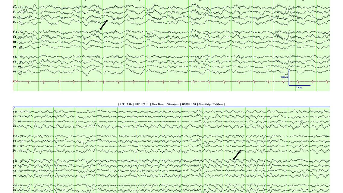
EEG results may be normal early in the disease course but as the encephalopathy worsens, generalized slowing, frequently in the delta range, which often becomes highly rhythmic, may be present (Figure). An EEG pattern termed “extreme delta brush” has been described in association with NMDA-R encephalitis.11
MRI of the brain may show T2 fluid-attenuated inversion recovery hyperintensity but is normal in the majority of individuals. Cerebrospinal fluid (CSF) typically shows pleocytosis. Screening for underlying malignancy is essential, with special attention for an ovarian teratoma in women.6

Figure. EEG (longitudinal bipolar montage) from a 30-year-old individual who presented with psychiatric symptoms and seizures, tested positive for NMDA-R antibodies, and was diagnosed with NMDA-R encephalitis. The EEG shows extreme delta brush, with the arrow indicating an example of a delta wave form with overlying fast activity (A). The EEG also demonstrates notched intermittent rhythmic monomorphic delta activity, with the arrow indicating an example of a notched delta wave form (B).
LGI1 Encephalitis
LGI1 encephalitis presents in individuals with a median age of 64 years and has a slight male predominance. Cognitive and memory issues, in addition to sleep disturbances and hyponatremia, may be present. About 90% of affected individuals have seizures.12 Faciobrachial dystonic seizures (FBDS; see the Video, available at www.practicalneurology.com), characterized by the sudden occurrence of a brief jerk of the arm and ipsilateral face, are highly suggestive of LGI1 encephalitis.13 These seizures typically occur multiple times per day and can be associated with brief altered awareness. Leg involvement may also occur, which can result in falls and associated injuries. Cognitive and memory decline are often present. FBDS are highly suggestive of LGI1 encephalitis but do not occur in all individuals. Other features that may suggest LGI1 encephalitis include a high seizure frequency, sometimes with multiple seizures per day, seizure brevity, ictal sensory symptoms, ictal piloerection, and provocation by hyperventilation.12,14
There is typically no EEG correlate during FBDS, although contralateral frontocentral infraslow discharges have been described preceding FBDS.15 EEG may also show temporal interictal epileptiform discharges, and subclinical temporal seizures may occur.12
MRI of the brain may show hippocampal and basal ganglia T1 or T2 hyperintensity but can be normal, as can CSF.16 Therefore, a high clinical index of suspicion is necessary to avoid missing this diagnosis. In contrast to NMDA-R encephalitis, antibody testing is more sensitive in serum than CSF for LGI1 encephalitis.16
GAD65 Antibody–Associated Seizures
High-titer GAD65 antibodies (defined as >20 nmol/L or >2000 U/mL in serum using radioimmunoassay and >10,000 IU/mL in serum or >100 IU/mL in CSF using enzyme-linked immunosorbent assay) are associated with diverse neurologic symptoms, including stiff-person syndrome, ataxia, vertigo, limbic encephalitis, and epilepsy.17 There can be overlap of each of these phenotypes. In addition to neurologic manifestations, type 1 diabetes, autoimmune thyroid disease, and pernicious anemia are also common in the setting of GAD65 autoimmunity.17
Individuals can present both with an acute or subacute limbic encephalitis presentation or with chronic temporal regional epilepsy.18,19 Temporal seizure semiology is common, including aura of déjà vu and symptoms suggestive of neocortical onset.10 Seizures provoked by music, known as musicogenic seizures, have been described in association with GAD65 antibodies.20
Bilateral temporal ictal and interictal manifestations are frequently present on EEG.
In limbic encephalitis presentations, MRI of the brain typically shows mesial temporal enlargement and T2 hyperintensity, sometimes progressing to hippocampal atrophy, although normal brain MRI results are not rare in individuals with chronic temporal lobe epilepsy presentations.19
Management
If seizures are linked to an autoimmune cause, or if acute encephalopathy suggests this may be the case, treatment typically involves immunotherapy. First-line immunotherapy typically includes corticosteroids, intravenous immunoglobulin, or both. Corticosteroids are typically given as high-dose intravenous methylprednisolone, or alternatively as high-dose oral steroid treatment, followed by tapering oral prednisone or weekly methylprednisolone infusions with a gradually increasing dose interval.21 Plasma exchange is also used acutely in severe cases. Depending on the clinical course, individuals at high risk for recurrence after initial treatments may be followed by a second-line approach using longer-acting immunosuppression, such as rituximab or cyclophosphamide.
Although many autoimmune-associated seizure disorders are monophasic, relapses may occur. Long-term immunotherapy with azathioprine, mycophenolate, or rituximab may be indicated in cases deemed to be at higher risk of relapse. Other immunotherapies with diverse mechanisms of action are being tested in clinical trials.
Response to immunotherapy depends on the underlying pathogenic antibody and mechanism and may be affected by delays in immunotherapy. Seizures associated with antibodies to cell surface antigens (including LGI1, NMDA-R, and others) typically have better response to immunotherapy, with many individuals having complete resolution of seizures. In one study of NMDA-R encephalitis, 88 participants had seizures at presentation, but all were seizure-free within 2 years.7 Resolution of seizures has been reported in 80% to 100% of individuals with LGI1 encephalitis after initiation of immunotherapy, which may occur within a few days after initiation.13,22
Seizures associated with antibodies against cell surface antigens have been conceptually categorized as acute symptomatic in nature rather than indicative of the establishment of epilepsy in this setting.23 In contrast, seizures in individuals with antibodies to intracellular antigens, including GAD65 antibodies and high-risk paraneoplastic onconeural antibodies, respond less favorably to immunotherapy and are more likely to develop autoimmune-associated epilepsy (Table 2).23
For individuals with high-risk paraneoplastic antibodies, screening for underlying malignancy should take place. Clinicians must be aware of certain tumor–antibody associations in individuals presenting with autoimmune-associated seizures. Small cell lung cancer may be associated with multiple antibodies, including GABAB receptor, ANNA1, AMPAR, and CRMP5 antibodies. Ovarian teratoma is associated with NMDA-R antibodies, and testicular cancer is associated with Ma2 and KLHL11 antibodies.24 Oncologic treatment is indicated for those with identified malignancies.
Antiseizure medications are typically used in conjunction with immunotherapy in individuals with autoimmune-associated seizure disorders. Seizure freedom has been reported in ~15% of individuals on antiseizure medication. Data on the efficacy of specific antiseizure medications are limited and studies are confounded by multiple factors, but sodium channel blocking medications appear to have greater efficacy compared with levetiracetam, especially in LGI1 encephalitis.22 A small study has suggested that clobazam and cenobamate may be efficacious in GAD65 antibody–associated epilepsy.25 However, drug-resistant epilepsy in this setting is common, occurring in ~75% of cases.18 Epilepsy surgery may be considered in some cases, but data are limited, and outcomes generally have been modest, with Engel I or II outcomes reported in 25% to 33% of cases.26-28

Conclusion
Autoimmune etiologies are important to recognize in individuals presenting with seizures given important management and prognostic implications. Certain clinical and seizure features may suggest a specific underlying antibody. Antibodies targeting NMDA-R, LGI1, or GAD65 are the most common in autoimmune-associated seizures and encephalitis. Immunotherapy is typically indicated when antibodies to cell surface antigens known to show immunotherapy responsivity are identified. When high-risk paraneoplastic antibodies are identified, malignancy screening is essential, as treatment of the inciting neoplasm in addition to immunotherapy is usually necessary to achieve optimal outcomes.
Ready to Claim Your Credits?
You have attempts to pass this post-test. Take your time and review carefully before submitting.
Good luck!
Recommended
 Epilepsy & Seizures
Epilepsy Essentials: Application of Magnetoencephalography in the Diagnosis and Treatment of Epilepsy
Abbas Babajani-Feremi, PhD; Giridhar P Kalamangalam, MD, DPhil, FRCPAbbas Babajani-Feremi, PhD; Giridhar P Kalamangalam, MD, DPhil, FRCP- Epilepsy & Seizures
Functional Neurologic Symptom Disorder Resolving After Endoscopic Encephalocele Repair
Charles F. Palmer, MD; Rodney J. Schlosser, MDCharles F. Palmer, MD; Rodney J. Schlosser, MD - Epilepsy & Seizures
Immunotherapy for Management of Refractory and Super-Refractory Status Epilepticus
Eugenia Ho, MD; Mackenzie Silverman, MDEugenia Ho, MD; Mackenzie Silverman, MD