
Epilepsy Essentials: Application of Magnetoencephalography in the Diagnosis and Treatment of Epilepsy
Used alongside MRI, video EEG, and other presurgical data, magnetoencephalography can localize epileptiform activity, guide invasive sampling, and support safer surgical planning in people with drug-resistant epilepsy.
KEY TAKEAWAYS
- MEG can add physiologic localization information when conventional presurgical epilepsy studies are inconclusive or discordant.
- Integrating MEG with structural imaging can help refine invasive monitoring strategies while identifying nearby eloquent cortex.
- New sensor technologies, network analysis, and AI-assisted approaches may further expand the role of MEG in individualized epilepsy evaluation.
Approximately 30% of people with epilepsy have drug-resistant seizures despite the availability of >25 antiseizure medications.1 For these individuals, resective surgery often offers a viable path to seizure freedom.2 However, identification of the appropriate surgical target can be challenging, particularly in MRI-negative (nonlesional) cases. Magnetoencephalography (MEG) provides a noninvasive approach to more precise identification of the epileptogenic zone by localizing the irritative zone—the region generating interictal epileptiform discharges—with millimeter-level spatial precision.3,4
A key advantage of MEG is that unlike surface EEG, in which scalp-recorded signals are distorted by the skull’s high resistivity, MEG is far less affected by conductivity differences, enabling more precise localization of neocortical sources.3 Thus, MEG can refine the epileptogenic hypothesis and narrow targets for further testing during presurgical evaluation of epilepsy, particularly when brain MRI, video EEG, neuropsychological assessment, PET, and other findings are inconclusive or discordant.5-7 Furthermore, MEG can guide phase II invasive monitoring with stereoelectroencephalography (SEEG) or subdural grids or strips while helping to identify eloquent cortex to support safer and more efficient surgical planning.5,6,8-10

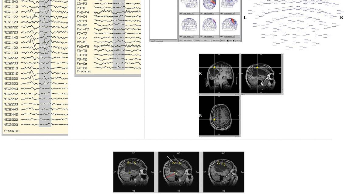
Figure 1. Magnetoencephalography (MEG) identifies additional epileptiform discharges adjacent to a large cavitary lesion. An individual with hemiplegic cerebral palsy, aged early 20 years, underwent presurgical evaluation for seizures characterized by behavioral arrest and incontinence, sometimes followed by hemiconvulsions. MRI showed a large right hemispheric cystic cavity. Scalp EEG demonstrated subtle right-sided and occasional right temporal spikes, whereas MEG localized frequent discharges along the superior parietal roof of the cyst, as well as a second cluster in the right mesial temporal lobe that was concordant with EEG findings. (A) Simultaneous MEG/EEG tracings show a representative interictal discharge clearly visible in MEG sensors but without a corresponding EEG spike. (B) Source analysis of the selected MEG spike, with the top left panel showing the spike time course from a highly sensitive MEG sensor, corresponding isofield maps, and estimated source; the top right panel showing time courses across all MEG sensors; and the bottom panel showing dipole localization at the selected time point. (C) Sagittal views of epileptiform discharge localizations. The white arrows indicate 2 frequent pericystic MEG-only discharge clusters that were not evident on scalp EEG during epilepsy monitoring unit evaluation. The red arrow marks the right mesial temporal cluster, which was concordant with the epilepsy monitoring unit EEG findings.
Clinical Role of MEG in Presurgical Epilepsy Evaluation
After failure of 2 appropriate antiseizure medication trials, individuals with drug-resistant epilepsy are ideally referred to an epilepsy center for presurgical evaluation.1,2 Typically, the latter involves epilepsy-protocol MRI, prolonged video EEG, neuropsychologic assessment, and selected adjunctive studies, such as MEG and PET.2,4 Within this framework, MEG is best viewed as an advanced physiologic localization tool that helps refine the anatomic-electroclinical hypothesis of seizure localization (Figure 1). In fact, MEG has 2 main contributions: localization of interictal epileptiform discharges and noninvasive functional mapping of eloquent cortex (Figure 2). Ictal, as opposed to interictal, MEG can provide additional localizing information, but its role remains limited by the difficulty of capturing seizures during relatively short recordings and by movement-related technical constraints.4 When MEG source estimates are projected onto the individual’s MRI, the result is termed magnetic source imaging, which allows spike sources to be interpreted in direct anatomic context. Beyond spike localization, MEG is used for functional mapping of language, motor, somatosensory, and other eloquent cortical regions when the suspected epileptogenic zone or planned resection is in the vicinity. This can help tailor SEEG electrode implantation and surgical planning while reducing the risk of postoperative deficit.5,6,11

Figure 2. Functional mapping with magnetoencephalography (MEG) for presurgical planning. (A) Somatosensory mapping in an individual with a left inferior parietal/supramarginal lesion, aged early 30 years, showed the hand area of the displaced left postcentral gyrus immediately anterior to the lesion. Blue markers indicate equivalent current dipoles corresponding to the P35m somatosensory evoked field; white arrows mark the lesion margin. (B) Motor mapping in an individual with a left parietal lesion, aged late 60 years, localized the motor cortex controlling the right hand immediately superior and anterior to the enhancing lesion. Orange markers indicate motor dipoles; white arrows mark the lesion margin. (C) Receptive language mapping in a right-handed individual with drug-resistant epilepsy, aged early 50 years, suggested left-hemisphere dominance for receptive language. (D) In the same individual, MEG spike localization demonstrated a cluster of frequent discharges in the left temporal lobe adjacent to receptive language cortex in the left hemisphere. Yellow triangles indicate dipoles associated with epileptiform discharges, red circles indicate receptive language sources, and blue squares indicate left somatosensory cortex.
When MEG Changes Clinical Decision Making
MEG most clearly changes management when the presurgical process moves from phase I hypothesis-building to phase II invasive sampling. Its clinical value extends beyond spike localization; MEG results can alter the placement of intracranial electrodes, the extent of the area requiring implantation, and occasionally, the surgical plan itself. In a prospective surgical planning study, MEG identified additional areas in 13% of participants, changed invasive EEG coverage in 23%, and changed the surgical decision in 20%. In a later evidence-based study of electromagnetic source imaging, source imaging provided clinically useful new information in 34% of participants,5,6 illustrating how MEG can become actionable rather than simply confirmatory.
MEG results are especially influential when the rest of the noninvasive workup is incomplete, partly discordant, or structurally unrevealing. In MRI-negative or subtle-lesion epilepsy, MEG can sharpen a lobar or sublobar hypothesis, prompt targeted MRI re-review, and help focus invasive monitoring on cortex that might otherwise remain unsampled.12,13 Recent reviews in MRI-negative focal epilepsy emphasize that MEG frequently adds localizing information that refines surgical planning, and multimodal studies show that MRI postprocessing and MEG findings are often synergistic rather than redundant.13,14 In MRI-negative focal epilepsy, morphometric MRI postprocessing may reveal subtle structural abnormalities in ~45% of individuals, and seizure freedom is more likely when the postprocessing-identified abnormalities are fully resected; these targets gain further support when they are concordant with magnetic source imaging.14
MEG also changes decision-making by adding prognostic information. Positive MEG findings are not all equivalent: individuals with a single tight cluster, stable dipole orientation, concordance with intracranial EEG results, and complete sampling or resection of the MEG-defined region have better postoperative seizure outcomes than those with scattered, bilateral, or incompletely sampled abnormalities.7,15,16 SEEG studies have shown that complete sampling of MEG clusters is associated with a markedly higher chance of seizure freedom, and outcome studies similarly show that concordant MEG and intracranial EEG localization and complete resection of MEG foci favor seizure freedom.15,16 At the same time, we caution against overinterpreting MEG findings. A negative or nonlocalizing MEG result does not exclude surgical candidacy, and MEG is less decisive when no interictal discharges are captured, abnormalities are broad or multifocal, or ictal MEG is limited by diffuse onset patterns or movement artifact.4,7,12,13
MEG results may also influence decisions in more complex scenarios, such as reoperations. In individuals with recurrent seizures after surgery, MEG may identify residual epileptogenic cortex near the resection margin, reveal an overlooked adjacent neocortical focus, or redirect reevaluation toward a broader network; several series support its role as a complementary test in failed-surgery workups.7,12,13 MEG may also help determine whether an individual is best served by additional targeted sampling, resection, or minimally invasive ablation. By contrast, its role in neuromodulation targeting remains promising but substantially less established than its role in SEEG planning or resective or ablative surgery. Current evidence is limited largely to conceptual reviews, case reports, and small pilot series, so this application is best regarded as emerging rather than standard practice.17
Applications of MEG in Pediatric Epilepsy: Opportunities and Practical Considerations
In pediatric drug-resistant epilepsy, MEG offers particular value because early and accurate localization can accelerate referral to surgery, preventing accumulation of developmental burden and taking advantage of greater neural plasticity in younger individuals.18 As in adults, MEG can be used to sharpen presurgical hypotheses when MRI findings are negative or subtle or when scalp EEG results appear generalized, multifocal, or discordant with imaging modalities.18 MEG-based functional mapping is particularly advantageous in children, in whom language and motor organization may be developmentally atypical or reorganized, making anatomy alone an unreliable guide.
MEG in children presents distinct challenges that require practical considerations. In younger children, smaller heads within adult-sized MEG helmets increase the sensor-to-cortex distance and allow greater head motion, both of which can reduce signal quality and localization accuracy; motion itself can blur the field pattern and increase source-estimation error. As a result, some children should be recorded while asleep or under sedation. Dexmedetomidine-based protocols appear feasible and may preserve interictal spike frequency better than alternatives, but sedation remains a tradeoff because it can alter spontaneous activity and limits active task mapping.19 Pediatric-specific helmets and emerging wearable optically pumped magnetometer (OPM)–based MEG systems may help reduce these barriers in the future.20-22
From Practical Constraints to Future Advances in MEG
MEG is highly informative when it captures well-formed interictal discharges, but such discharges do not occur in every individual. Routine clinical standards call for at least 30 minutes (typically 1 to 2 hours) of spontaneous MEG–EEG recording, ideally including both wakefulness and sleep, yet some studies remain nondiagnostic simply because no habitual epileptiform activity is recorded. Ictal MEG is even more limited, largely because seizures are rarer and the data are degraded by motion and other technical constraints.
MEG also has certain anatomic and physiologic limitations. In general, MEG is less sensitive to deep or radially oriented generators,3 and source localization is less reliable when epileptic activity is spatially extended or rapidly propagates from the generating area.23 In addition, standard dipole models are strictly applicable only when a limited cortical region is synchronously active; widespread or unstable source patterns should be interpreted cautiously. In such circumstances—when the goal is to estimate source extent, onset propagation, or localization of noisy, spatially extended, or complex events—the dipole fit should be complemented by distributed source modeling (such as minimum norm or standardized low-resolution brain electromagnetic tomography [sLORETA]), or, for the specific event under study, replaced by distributed source modeling as the primary localization approach.24
One important but underemphasized issue concerns the procedural standardization of MEG across centers. For example, in presurgical language mapping, practice remains heterogeneous in task selection, stimulus design, analysis pipelines, source modeling, laterality metrics, and reporting.10,25 Greater harmonization is essential for robust multicenter validation and broader clinical adoption.
More recently, MEG has seen a period of rapid innovation driven by wearable sensors, network-based biomarkers, and artificial intelligence–assisted analysis. Together with tighter multimodal integration, these advances may expand MEG beyond conventional single-session presurgical localization toward more accessible, motion-tolerant, and individualized evaluation. A major hardware advancement is OPM-based MEG. Because OPM sensors can be placed on or near the scalp and do not require cryogenic dewars, they reduce sensor-to-cortex distance and better accommodate head movement. This makes OPM-based MEG especially attractive for use in children or individuals with hyperkinetic movement disorders. Early studies suggest that higher-density OPM systems can approach or, in some settings, surpass the performance of conventional superconducting quantum interference device (SQUID)–based MEG systems, but routine clinical adoption will require robust shielding or field control, calibration, interference suppression, and regulatory validation.20-22
Another major direction is the shift from lesion-centered localization to network-level characterization. In epilepsy, MEG connectivity and graph theory analyses are being explored to define epileptic networks and seizure propagation architecture.26 Recent studies support strong test–retest reliability and heritability of resting-state MEG network measures, highlighting their potential as biomarkers.27,28 However, large multicenter validation studies are lacking, and further work is needed before these measures can be translated into routine clinical biomarkers for presurgical evaluation in epilepsy.
Summary
MEG is best viewed as a high-value adjunct within the comprehensive presurgical evaluation of drug-resistant epilepsy, where it adds direct noninvasive physiologic information that can sharpen the epileptogenic hypothesis, improve confidence in localization, guide SEEG or selected grid or strip implantation, and help delineate eloquent cortex when resection or ablation is being considered. It is particularly valuable in MRI-negative, subtle-lesion, neocortical, multilobar, pediatric, and other discordant cases, where it can transform an imprecise phase I evaluation into a more focused, testable, and surgically actionable plan.
At the same time, MEG should be used thoughtfully and interpreted in context. Its yield depends on capturing analyzable epileptiform activity, and its sensitivity is lower for deep or broadly distributed generators than for focal neocortical sources; thus, a negative or nonlocalizing study does not exclude surgical candidacy.
The field of MEG is evolving. Wearable OPM-based systems, network-level analyses, and artificial intelligence–assisted workflows may make MEG more accessible, motion-tolerant, and informative, particularly among children and other challenging populations. Broader clinical translation will require stronger cross-center standardization and multicenter validation. In that sense, MEG remains both a clinically actionable tool in today’s epilepsy practice and a platform for future advances in individualized presurgical decision-making.
Ready to Claim Your Credits?
You have attempts to pass this post-test. Take your time and review carefully before submitting.
Good luck!
Recommended
 Epilepsy & Seizures
Machine Learning–Based Neuroimaging in Epilepsy: Improving Detection of Epileptogenic Lesions
Justyna Ekert, MBBS, PhD; Erik H. Middlebrooks, MDJustyna Ekert, MBBS, PhD; Erik H. Middlebrooks, MD- Epilepsy & Seizures
Immunotherapy for Management of Refractory and Super-Refractory Status Epilepticus
Eugenia Ho, MD; Mackenzie Silverman, MDEugenia Ho, MD; Mackenzie Silverman, MD