Portable Brain Imaging System Shows Promise for Stroke Identification
Preliminary results have been released from a clinical trial comparing stroke imaging with a portable electromagnetic variation (EMV) device (EMVision, Brisbane, Australia) to stroke imaging with CT and MR fluid attenuated inversion recovery (FLAIR). An imaging algorithm team received blinded datasets and found correlation between EMV and CT or MR FLAIR including detection and localization of abnormal brain tissue.
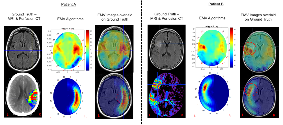
These initial datasets have been reviewed extensively by EMV clinical advisors, including neurology and radiology experts. The EMV functional imaging scans demonstrate a strong correlation with the ground truth scans for these participants (MRI and CT) (Figure). This is the first set of fully analysed data with complete clinical assessment.
The EMV device comprises a lightweight headset with an array of antennas that transmit safe low-power electromagnetic signals into the brain. Software is used to compare the scatter of the electromagnetic energy and create an image displaying variation between healthy and unhealthy brain tissue. The device is fully portable, weighing approximately 6 pounds, making it usable in the prehospital setting or at the bedside.
The EMV scans shown in the Figure were taken after initial treatment and “ground truth” MRI FLAIR and CT perfusion scans were obtained. The red regions in the EMV image represent higher contrast in electrical properties, as is typical of anomalies. Both participant cases involved a reasonably small infarct (visible on the MRI FLAIR images) and a larger penumbra. For both cases, the technique produces accurate localization of abnormal brain tissue which is clearly distinguished from the surrounding brain tissue. This is typically more difficult on plain CT scans.
Cochairs of the Australian Stroke Alliance and past presidents of the World Stroke Organization, professors Stephen Davis AM and Geoffrey Donnan AO also provided expert feedback. Professor Davis commented, “These early images are clinically promising, clearly showing the effects of ischemic stroke in the same region as the gold standard imaging methods”. Donnan commented, “the lightweight portability of the device makes it a potential candidate for emergency stroke imaging in the prehospital setting.”
EMVision chief executive officer, Dr. Ron Weinberger, commented “Our first set of images, while preliminary, is certainly encouraging, demonstrating a strong correlation with mainstay medical imaging outputs, with the potential to add unique functional information. We are confident that as we continue to process further stroke patient data, we will demonstrate our unique value proposition to meet a major unmet clinical need in rapid and portable stroke diagnosis and monitoring.”